




















AI Detects Brain Lesions MRI Could Not See
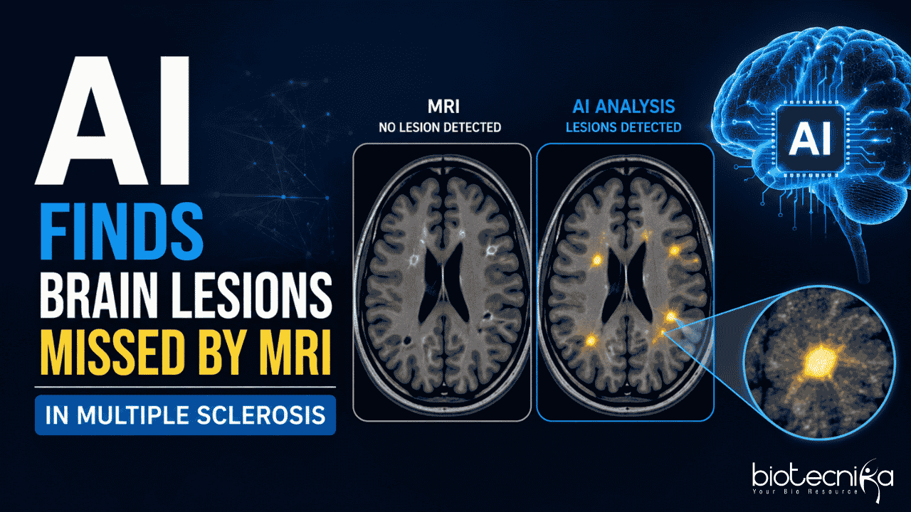
Artificial Intelligence (AI) in healthcare has improved how diseases are diagnosed and treated. Integration of AI in disease management has uncovered the hidden patterns, improving the treatment efficiency. One such breakthrough in neuroscience is the detection of brain lesions in people suffering from multiple sclerosis. AI helps doctors detect lesions in the gray matter, which is not possible with regular MRI. They believe this could help them understand the disease better and find different treatment methods.
Why Do These Brain Lesions Matter
Multiple sclerosis is an autoimmune neurological disorder in which the immune system fails to recognise its own body cells and attacks the myelin (the protective sheath of nerve fibres). Multiple sclerosis affects both the white matter and the cortical region (the outer surface of the brain). Unfortunately, the detection of the cortical lesions was not considered due to limitations in the MRI. MRI scans can easily capture the lesions in the white matter, but not the lesions in the gray matter, which in turn affects the treatment progression.
For years, doctors have relied on MRI scans to track the disease by looking for lesions in the brain’s white matter. Though researchers and doctors have known about the involvement of cortical lesions in the gray matter since the 19th century, due to the clinical limitations of MRI scans, they weren’t included as part of the diagnosis. Hence, an important part of the disease is largely hidden from view.
How AI Helped Reveal the Invisible
To take the research further, Scientists from the University at Buffalo and collaborating institutions developed an AI-assisted approach. This novel method analyses multiple MRI images together rather than examining each image separately. The system looks for subtle patterns and differences across scans that are too small for the human eye to notice.
“The hallmark of the disease is focal demyelinating lesions, and for ages, only white matter was considered with respect to multiple sclerosis,” says the team.
The team combined several advanced image-processing methods, including a newly developed technique called Multimodal Cortical Lesion Enhancement (MMCLE). These tools were applied to MRI data from the Phase III ORATORIO clinical trial, which included more than 700 participants with multiple sclerosis. This was an FDA regulatory study of Ocrelizumab, a multiple sclerosis drug.
Robert Zivadinov, MD, PhD, highlights the importance of this AI- assisted approach. Finding cortical lesions, which was once an impossible task in MRI, is made possible by the latest technology. “Being able to visualise one of the important factors of the disease progression for the first time is indeed a major advancement in neuroscience, and this helps in understanding the cognitive impairment and dysfunction related to multiple sclerosis,” he adds.
It is also found that the cortical lesions or the gray matter lesions are seen in the early stages of the disease progression. This proves that gray matter pathology is not something to be neglected and is equally important as white matter damage.
Thousands of Hidden Lesions Detected
Yes, they developed MMCLE, but what are the findings? The results were striking. By default, the conventional MRI reports showed white matter lesions, while the enhanced AI-mediated image analysis revealed 15 to 20 cortical lesions per patient. The researchers were able to identify more than 11,000 such lesions, which went undetected previously, across the entire dataset.
With these methods, the cortical lesions are much easier not only to detect but also to quantify. The team confirmed that using AI and Deep learning, the simultaneous use would dramatically improve the quantification.
This means a lot to the researchers, as the technologically advanced findings provide a clear vision of the disease development. The significant amount of brain damage that went unnoticed over decades has finally been understood and proven. The traditional MRI, along with AI assistance, creates a new revolution in the way multiple sclerosis is diagnosed.
What This Means for Multiple Sclerosis Research
This tells the world that even the most advanced scanning couldn’t reveal everything that is happening inside the body. At the same time, tagging the advanced technologies with the existing pipelines could improve healthcare monitoring a lot. This means there’s a chance to revisit the previous finding or clinical trials and explore more about what’s hidden. Researchers believe the technology could help evaluate older treatments more accurately and guide the development of future therapies.
As AI continues to advance in medical imaging, this approach could offer clinicians a powerful new tool for tracking multiple sclerosis. By bringing hidden brain lesions into view, researchers hope to gain deeper insights into the disease and improve outcomes for patients worldwide.