


Scientists Use New Imaging Modality to Detect Cancer in Tissues
Monitoring biomarkers and injected theranostic nanomaterials in tissues and organs plays a pivotal role in numerous medical applications ranging from cancer diagnostics to drug delivery. Scanning electrochemical microscopy has been demonstrated as a powerful tool to create highly resolved maps of the distributions of relevant biomolecules in cells and tissues without suffering from the optical interferences of conventional microscopy.
In the field of theranostics – a portmanteau of the words “therapy” and “diagnostics” – researchers use spatial information about cancer cells in the body to come up with targeted therapies. But this approach requires fluorescent markers. Electrochemical imaging, which uses only the tissue’s natural – or endogenous – electrochemical markers, could also provide doctors with what they need to make diagnoses and prescribe therapies.
Now, researchers at the École polytechnique fédérale de Lausanne (EPFL) in Switzerland and Linkou Chang Gung Memorial Hospital in Taiwan have developed a way of imaging biochemicals in the body using electric current. The technique, named scanning electrochemical microscopy, has already been used to map out hemoglobin throughout a heart of a mouse.
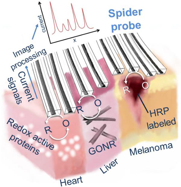
This device has eight flexible microelectrodes that are lined up side by side. The device scans tissues immersed in an electrolyte solution in order to measure the electrochemical response. Some types of molecules and nanomaterials that have built up in the tissue exchange electrons with the electrochemical mediator in the solution, and the resulting electric currents are used by the researchers to reconstruct the image.
The soft microelectrodes are brushed gently across the tissue samples, while they measure the electrical current produced by certain chemicals in the tissues to get an idea of the physical structure of that tissue as well as its composition.
The device then delivers a variety of electrical signals of different frequencies and amplitudes into the object’s tissue while measuring the response. When certain biomolecules begin to exchange electrons with the electrolyte, the overall charge of the solution changes and this can be detected. Because these events happen at known concentrations and energy levels, the system can provide the location and nature of the electrical discharges. This is rendered into a unique image using a computer. Pretty nifty and is essentially a new imaging modality that doesn’t require contrast agents nor ionizing radiation.
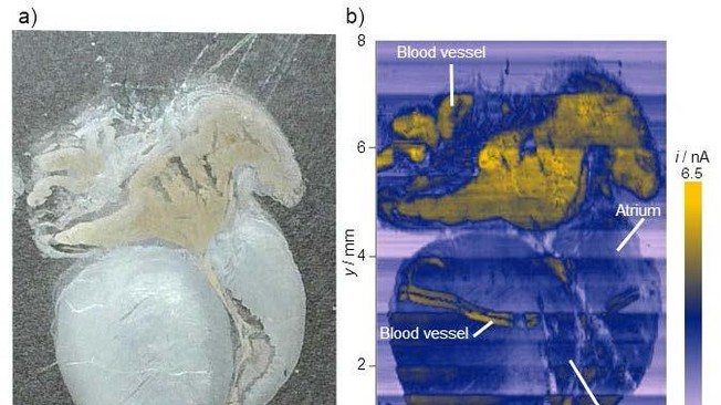
The team provided three separate demonstrations of this technique’s use. In the first, it scanned mouse livers to show that a certain type of nanoribbon that’s being studied as a potential drug delivery mechanism can be distributed throughout the liver. In the second, the probes measured hemoglobin proteins to get a full image of a mouse heart (the right side of the image above). And in another experiment, the researchers used the technique to show that it can accurately differentiate healthy human tissue from cancerous tissue.
“We are perfectly capable of using electrochemistry to kill cancer cells on microscope slides and in petri dishes, but doing so in thick tissue is another story,” says Hubert Girault, who runs the Laboratory of Physical and Analytical Electrochemistry. Indeed, Professor Girault hopes that it will one day be possible to use electrochemical microscopy during surgery. He envisions a device with interconnected microelectrodes capable of producing an image that will test for tumors and then electrochemically destroying any cancerous cells found by applying a burst of voltage: “Around two volts, that’s not much, but it’s enough to generate oxygen radicals and eliminate cancer cells,” says Girault.


