


CSIR NET UNIT 7 Notes- Cardiovascular System
While this unit may seem quite challenging and lengthy, students with zoology, physiology, or medical background can make this unit a scoring advantage. Students from other educational backgrounds can also prepare for particular topics from this section of UNIT 7B – Cardiovascular System – related to which questions are often asked in the CSIR NET exam.
Reference Books FOR CSIR NET UNIT 7:
- Principles of anatomy physiology by Gerard J. Tortora / Bryan Derrickson
- Principles of Animal Physiology by C. D. Moyes and P. M. Schulte
- Schaum’s Outline of Human Anatomy and Physiology
- Guyton and Hall Textbook of Medical Physiology (Guyton Physiology) by John E. Hall
CSIR NET UNIT 7 Notes – 7B : Cardiovascular System
Introduction:
- The cardiovascular system consists of the heart and blood vessels.
- Sends blood to lungs for oxygen to reach body cells and exchange with them. materials
- Also circulates waste products to specific organ systems for removal from the blood.
- The heart beats about 100,000 times every day.
Amphibians and reptiles
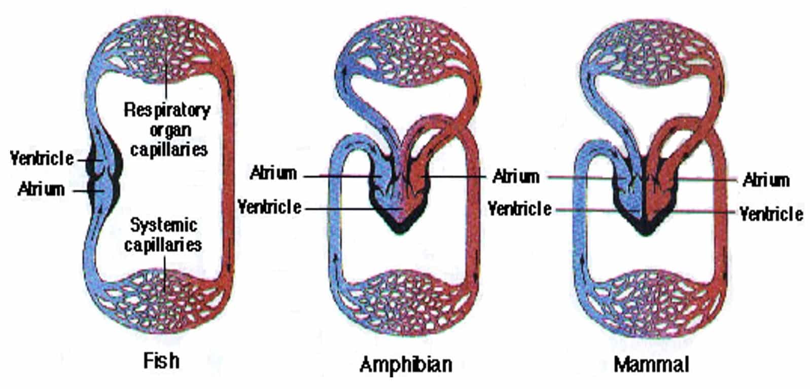
Fig: Blood circulation – Single loop fish (2 chambered) and double loop amphibian (3 chambered) and mammal (4 chambered) circulation.
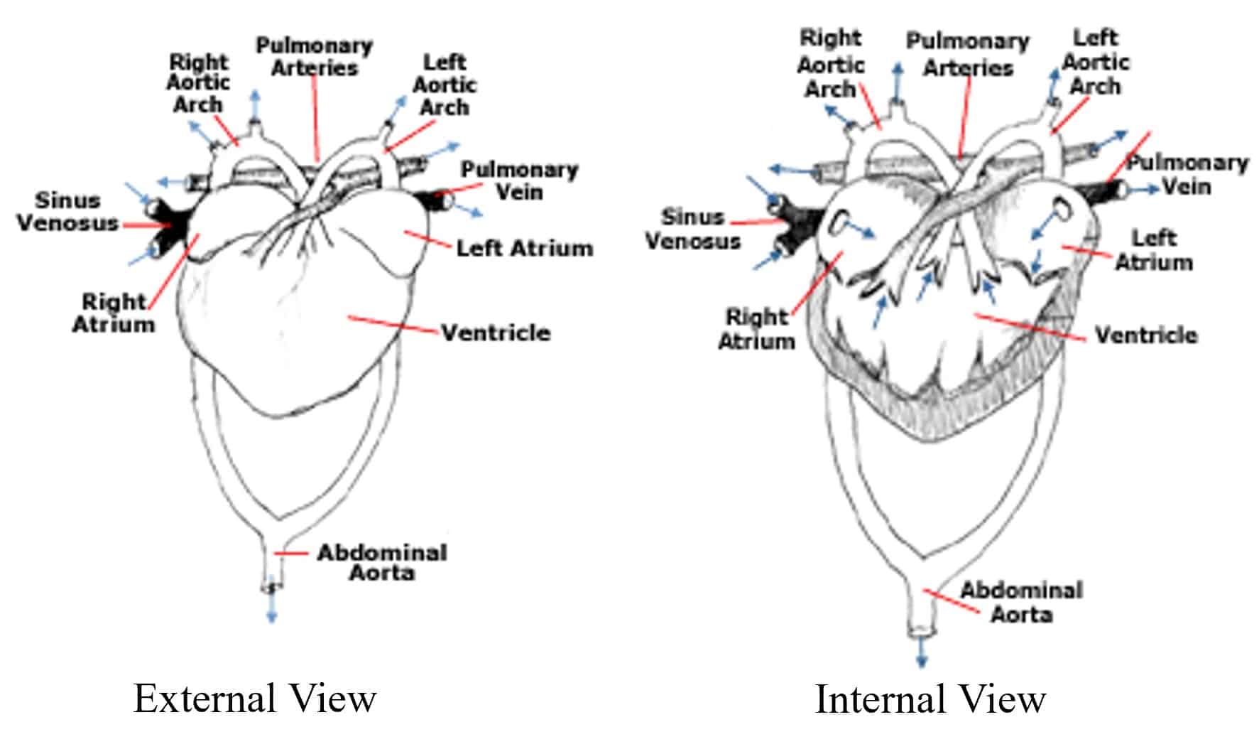
Fig: Amphibians & Reptiles Heart – External & Interior View
- Amphibians and reptiles have a three-chambered heart.
- The three-chambered heart consists of two atria and one ventricle. (The crocodile is said to have a four-chambered heart.)
- Blood leaving the ventricle passes into one of two vessels.
- It either travels through the pulmonary arteries leading to the lungs or through a forked aorta leading to the rest of the body.
- Oxygenated blood returning to the heart from the lungs through the pulmonary vein passes into the left atrium, while deoxygenated blood returning from the body through the sinus venosus passes into the right atrium.
Mammals and Birds
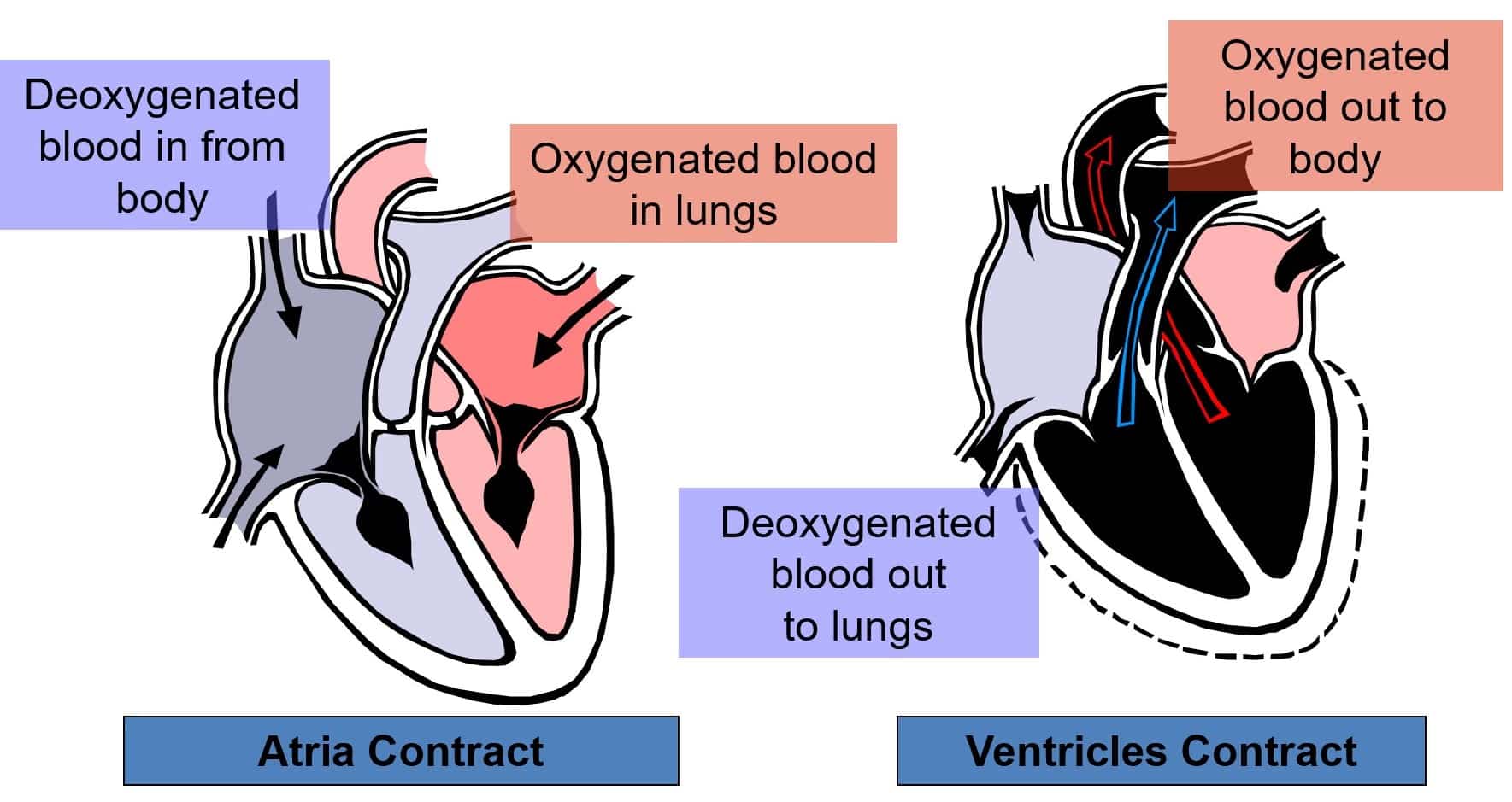
- Mammalian and avian hearts have four chambers – two atria and two ventricles.
- The most efficient systems are not mixed as deoxygenated and oxygenated blood.
- The right atrium receives deoxygenated blood from the body through the inferior and superior vena cava.
- The blood then passes to the right ventricle to be pumped through the pulmonary arteries to the lungs, where it becomes oxygenated.
- It returns to the left atrium via the pulmonary veins; this oxygen-rich blood is then passed to the left ventricle and pumped through the aorta to the rest of the body.
- The aorta is the largest artery with enormous stretch and elasticity to withstand the pressure created by the pumping ventricle. (CSIR UNIT 7 Notes by Biotecnika)
Structure of the Heart
- Cone-shaped organ about the size of a loose fist

- In the mediastinum
- Extends from the level of the second rib to about the level of the sixth rib
- Slightly left of the midline
- The heart is bordered:
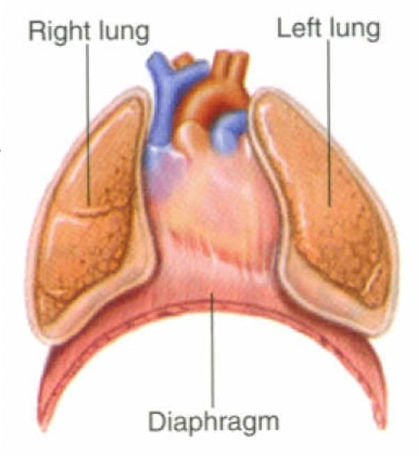
-Laterally by the lungs
-Posteriorly by the vertebral column
-Anteriorly by the sternum - Rests on the diaphragm
PERICARDIUM
- The pericardium
(1) the fibrous pericardium and
(2) the serous pericardium - The superficial fibrous pericardium is composed of tough, inelastic, dense irregular connective tissue, prevents overstretching of the heart, provides protection, and anchors the heart in the mediastinum.
- The deeper serous pericardium is a thinner, more delicate membrane that forms a double layer around the heart.
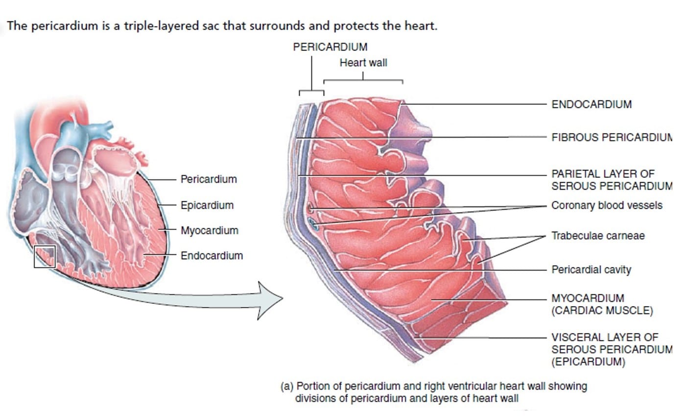
Fig: The pericardium & Heart Wall
- The outer parietal layer of the serous pericardium is fused to the fibrous pericardium. The inner visceral layer of the serous pericardium is called the epicardium.
- Between the parietal and visceral layers of the serous pericardium is a thin film of lubricating serous fluid.
- This slippery secretion of the pericardial cells, known as pericardial fluid, reduces friction between the layers of the serous pericardium as the heart moves.
CSIR UNIT 7 Notes
Structures of the Heart (cont.)
Four chambers
- Two atria

-Upper chambers
-Left and right
-Separated by the interatrial septum - Two ventricles
-Lower chambers
-Left and right
-Separated by the interventricular septum - The atrioventricular septum separates the atria from the ventricles
- On the anterior surface of each atrium is a wrinkled pouchlike structure called an auricle.
- Each auricle slightly increases the capacity of an atrium to hold a greater volume of blood.
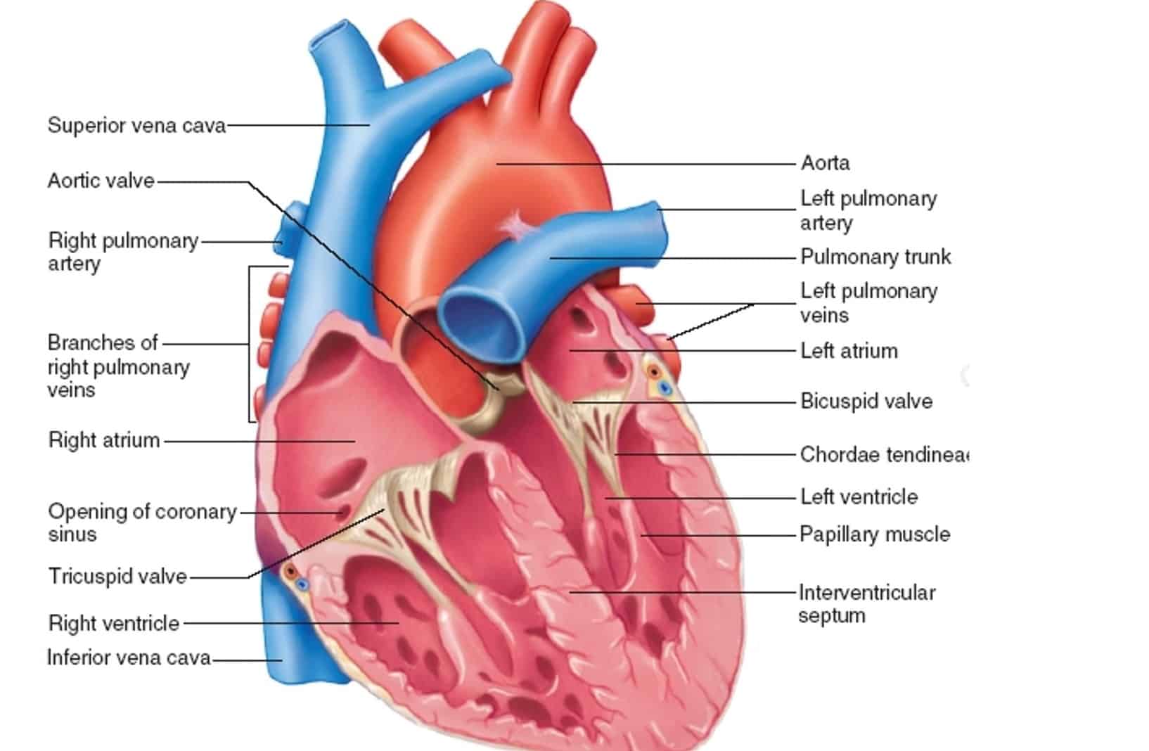
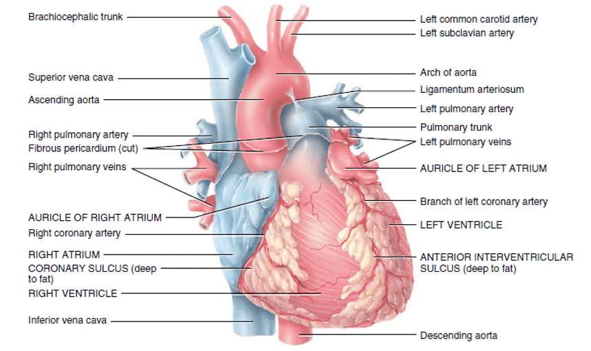
Fig: The surface of the Heart: Surface Features
CSIR UNIT 7 Notes by Biotecnika
Download CSIR UNIT 13(A) Notes On Gene Cloning, rDNA Technology – Methods in Biology.
- The surface of the heart- is a series of grooves, called sulci, that contain coronary blood vessels and a variable amount of fat.
- Each sulcus marks the external boundary between two chambers of the heart.
- The deep coronary sulcus encircles most of the heart and marks the external boundary between the superior atria and inferior ventricles.
- The anterior interventricular is a shallow groove on the anterior surface of the heart that marks the external boundary between the right and left ventricles on the anterior aspect of the heart.
- Tricuspid valve – prevents blood from flowing back into the right atrium when the right ventricle contracts.
- Bicuspid (mitral) valve – prevents blood from flowing back into the left atrium when the left ventricle contracts.
- Pulmonary semilunar valve – prevents blood from flowing back into the right ventricle.
- Aortic semilunar valve – prevents blood from flowing back into the left ventricle.
Cardiac Muscle Histology
- The cardiac muscle fiber is 50–100 µm long and has a diameter of about 14µm.
- The ends of cardiac muscle fibers connect to neighboring fibers by irregular transverse thickenings of the sarcolemma called intercalated discs.
- The discs contain desmosomes, which hold the fibers together, and gap junctions, which allow muscle action potentials to conduct from one muscle fiber to its neighbors.
- Gap junctions allow the entire myocardium of the atria or the ventricles to contract as a single, coordinated unit.
CARDIAC CYCLE – CONTD…
Download CSIR UNIT 7 FULL Notes On Cardiovascular System
Launching CSIR NET Raftaar Batch 2022: Speed Revision Batch for CSIR NET September 2020 Exam
As the CSIR exam approaches, Biotecnika launches “Raftaar CSIR NET Batch” to help all the CSIR aspirants with a quick and effective learning program to strengthen their preparation and make them exam-ready. This will be a speed revision batch where our CSIR NET Exam experts will train you on tackling and revising for the upcoming exam. Do not miss out on the excellent pre-exam preparative Raftaar CSIR NET Course. Limited Seats are available.
START DATE: 19th July 2022
Why Raftaar Batch for CSIR NET?
Let’s face the fact that we are all out of touch with the CSIR NET Exam( Thanks to the long gap). To help the students prepare for the Upcoming Exam, we designed a perfect Raftaar CSIR NET course. Here you will get to interact with experts, revise important topics and get plenty of additional facilities to aid you in the last 60 days of exam preparation.


 2026 Notification Released – Check Eligibility, Syllabus & Apply Online")


