Microscope Images Of Coronavirus Captured For The First Time By NIH
The first images of the deadly coronavirus have been released by US scientists. The coronavirus that originated in the city of Wuhan in China during late December 2019 has killed around 1,700 people and infected more than 70,000 people around the world.
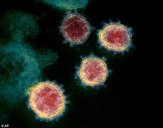
The National Institute of Health Scientists collected the samples from an infected person and captured the images of viruses emerging from various cell types using an electron microscope.
As expected, the microscope images of new coronavirus shared some similarities with that of SARS, as the name SARS-CoV-2 of new coronavirus indicates.
Viruses are different in structure from bacteria or any other living cell. They consist of either RNA or DNA enclosed in a protein shell called the capsid. One virus differs from the other in terms of its genetic makeup, DNA or RNA and the proteins on the exterior that help the virus to invade other cells.
Coronavirus has spiked protein shells that give them the resemblance of a crown or corona, which gave them the name. The microscopic images released by NIH shows these spike proteins as slightly fuzzy points protruding from the exterior of each virus particle.
NIH scientists Vincent Munster and Michael Letko sequenced the spike proteins of the virus and identified its close resemblance with SARS.
The resemblance with SARS made the World Health Organization (WHO) name the virus as SARS-CoV-2 and the disease as COVID-19. They also created a naming convention that links the two viruses together.
The analysis of proteins that make up the spiked shell also revealed the original source of the virus as bats. But what makes the virus able to penetrate human cells is the mutations that happened to them along their evolutionary progression.
The microscope images of coronavirus show the viruses emerging from some cells to go attack other cells. Sometimes, they can be seen in clusters also.
Viruses are so tiny that they can’t be seen with light microscopes. Scientists use high-powered electron microscopes to see the viruses. And the images from the electron microscope showed the coronaviruses moving between cells to feed off.
Identification of the viral structure could give scientists clues about how to disarm them. Currently, Chinese scientists are giving antiviral drugs and plasma transfusions (which contain antibodies the immune system built to fight the new coronavirus) from those who recovered from the disease to the infected individuals.
Trials are underway to use antivirals for Ebola and HIV antivirals to treat the disease. The ebola antivirals seem to be working on macaque monkeys affected with MERS, another coronavirus in the family. It could be used to treat and prevent COVID-19 as both MERS and SARS-CoV-2 share similar protein spikes.
And its these genomic and structural similarities that suggest the vaccine that US scientists developed to treat SARS during the 2003 outbreak and have now taken out of storage might be modifiable to work against the new virus that’s raced around the globe and infected more than 71,000 people.