


")


Where Life Begins Again: The Lab-Grown Mini Amniotic Sac Behind a Silent Scientific Revolution
A significant step was taken towards Regenerative Science and Developmental Biology by some renowned Researchers of the prestigious ‘Francis Crick Institute’ in London. The Research team has successfully grown ‘amniotic sacs’ miniatures in a cultured and conditioned laboratory utilizing human Embryonic Stem Cells (ES).
These miniature Amniotic Sac structures replicate the early developmental stages of the human Amniotic Sac, which generally forms during the first month of pregnancy. This incredible Research provides an opportunity for Scientific professionals to explore and understand the ‘early stages’ of human embryo development. Generally, this early stage has been a challenge to access worldwide due to technical and ethical limitations in the Scientific field.
The Research was published in the prestigious journal “Cell” on July 10, 2025. It introduces PGAs (Post-Gastrulation Amnioids), which are 3D (three-dimensional) models that replicate the natural amniotic sac’s structure and remain viable for over three months in a regulated laboratory culture.
The senior author of the Research, Dr. Silvia Santos, said, “I’m extremely excited about the potential of these little structures.” She further stated, “We’re finally able to study a part of development that has remained hidden for so long.”
Importance of the Amniotic Sacs
The Amniotic Sacs form right after fertilization, surrounding the growing embryo in the body. It acts like a fluid-filled cushion, providing protection, nourishment, and support to the growing fetus during the early stages of pregnancy development. The amniotic sac, along with other supporting tissues as well as the placenta, plays essential roles in sustaining and growing life in the womb during pregnancy.
To support this notion, Dr. Santos further explained, “Supporting tissues like the placenta, like the amniotic sac, grow with the embryo and are important for the embryo’s growth and survival.”
The amniotic sac is one of the components of early pregnancy that has been less explored, possibly since the first few weeks of pregnancy are pretty challenging to study and Research in human beings.
Earlier laboratory models have generally failed to capture the amniotic sac’s complex, a two-layered structure. The main reason could be that it typically survives for a few days, which in turn limits the understanding of sustained gestation developmental processes.
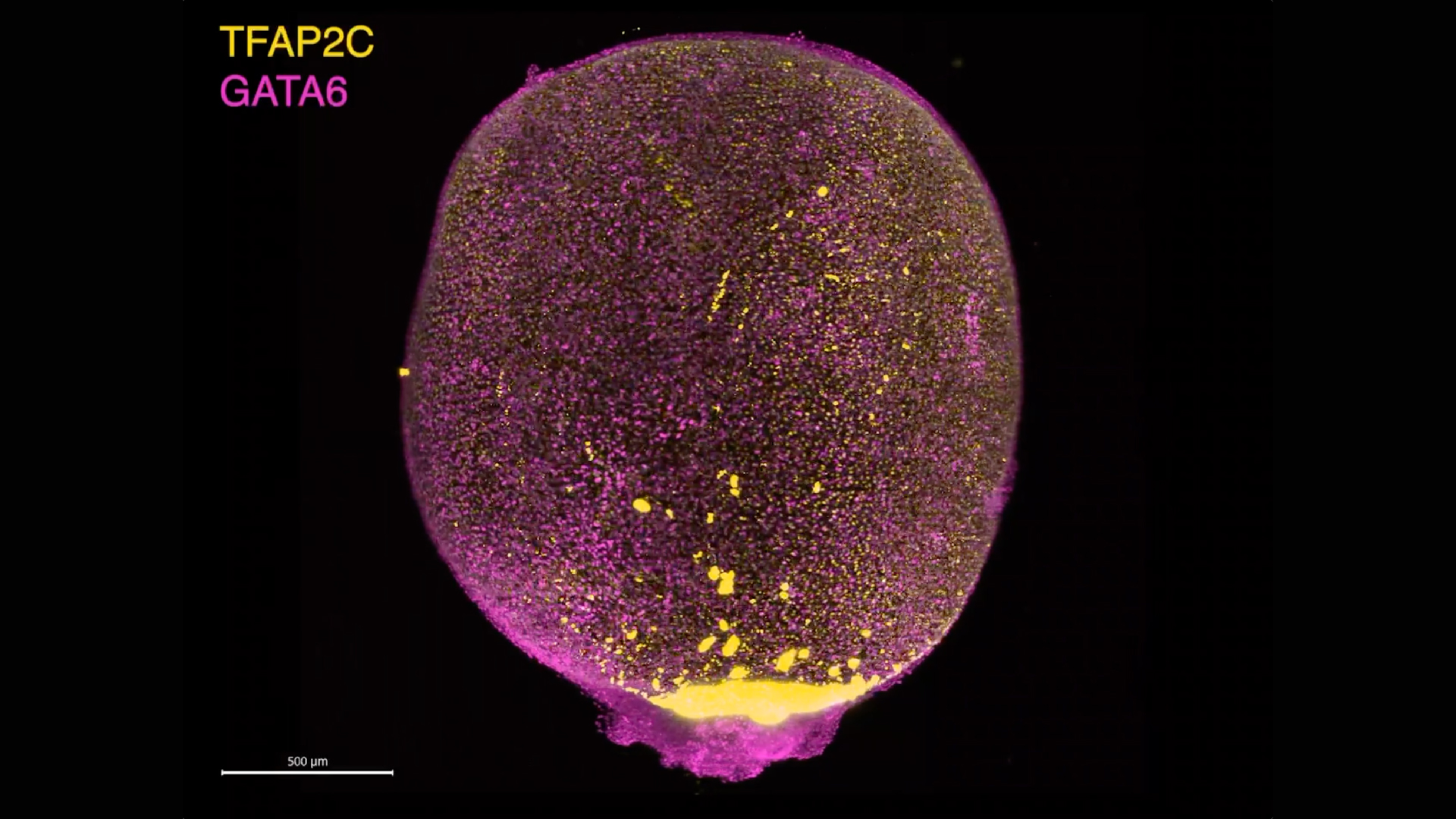
This image shows one of the new lab models, called PGAs, at day 18 of growth. PGAs are composed of two different cell populations (magenta, the extra-embryonic mesoderm and yellow, the amniotic endoderm). (Image credit: Gharibi, B. (2025). Cell.)
A Self-Organizing Achievement
To address these limitations, the Research team developed a laboratory culture method utilizing Human Embryonic Stem Cells (hES), which have the capability to create and differentiate into any cell type in the human body. They exposed the Stem Cells to two major signaling molecules, namely CHIR and BMP4, for over a 48-hour time period.
First day BMP4 signalling molecule was conditioned for 24 hours, which was followed by CHIR signalling molecule for the next day.
Dr. Santos further explained that they left the cells alone in round-bottomed culture dishes. “The rest was complete self-organization,” which emphasized that the maturing Stem Cells organized their own assembly into the structure. Once the period was over, the Research team transferred the cells into round-bottomed culture dishes and let them develop with time.
The Research outcomes were astonishing. The cells were fluid-filled sacs, formed a spherical shape, and aggregated spontaneously, with the same two-layered structure as witnessed in natural human amniotic sacs.
Dr. Santos stated, “This just shows you that these embryonic stem cells have this amazing propensity to specialize and to become everything given the right instructions, which I’m still in awe about.”
Furthermore, the PGAs had grown around 2.5 centimeters approximately in size, which is equivalent to a real human amniotic sac at around first month of pregnancy.
She further stated to “Live Science” that, “They are little golf balls,” And explained that the PGAs also form the Lab-Grown Amniotic Sac’s distinct two-layer structure.
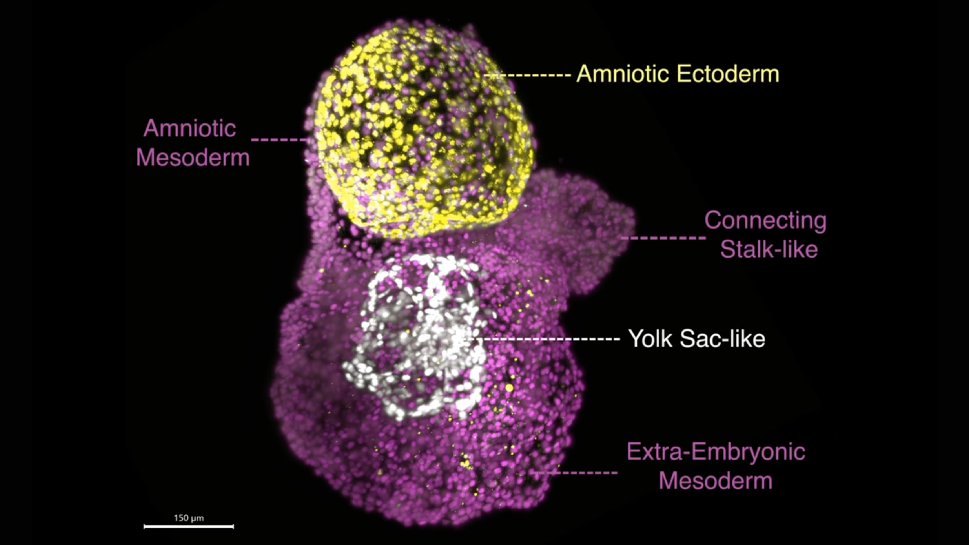
Fig.: This image shows a PGA at day 8 of growth, displaying the early tissues that support the embryo, including the amniotic sac and the yolk sac-like structures, surrounded by extraembryonic mesoderm tissue. (Image credit: Gharibi, B. (2025). Cell.)
One gene but with a significant influence
With the successful formation of Post-Gastrulation Amnioids, the researchers had identified which particular Genes could be responsible for triggering his transformation. After systematically interfering with a range of genes thought to influence early development, the scientist has discovered a standout regulator, namely GATA3.
A transcription factor, GATA3, converts Stem Cells into amniotic-like cells by itself, with no requirement for chemicals in addition. This factor also regulates molecules as well as BMP4, which are used in the original Research Culture Protocol. This study suggested that “GATA3” acts like a master switch, which generates signals needed for the formation of Amniotic Sacs.
Apart from forming on their own, the PGAs also explain the ability to influence their cellular environment. When the researchers placed fresh, undifferentiated stem cells next to the Post-Gastrulation Amnioid, those neighboring cells began to transform into extraembryonic lineages, just like those found in the yolk sac and placenta.
This research confirms that the amniotic sac serves as an active signaling hub, helping to guide nearby cells during early embryonic development.
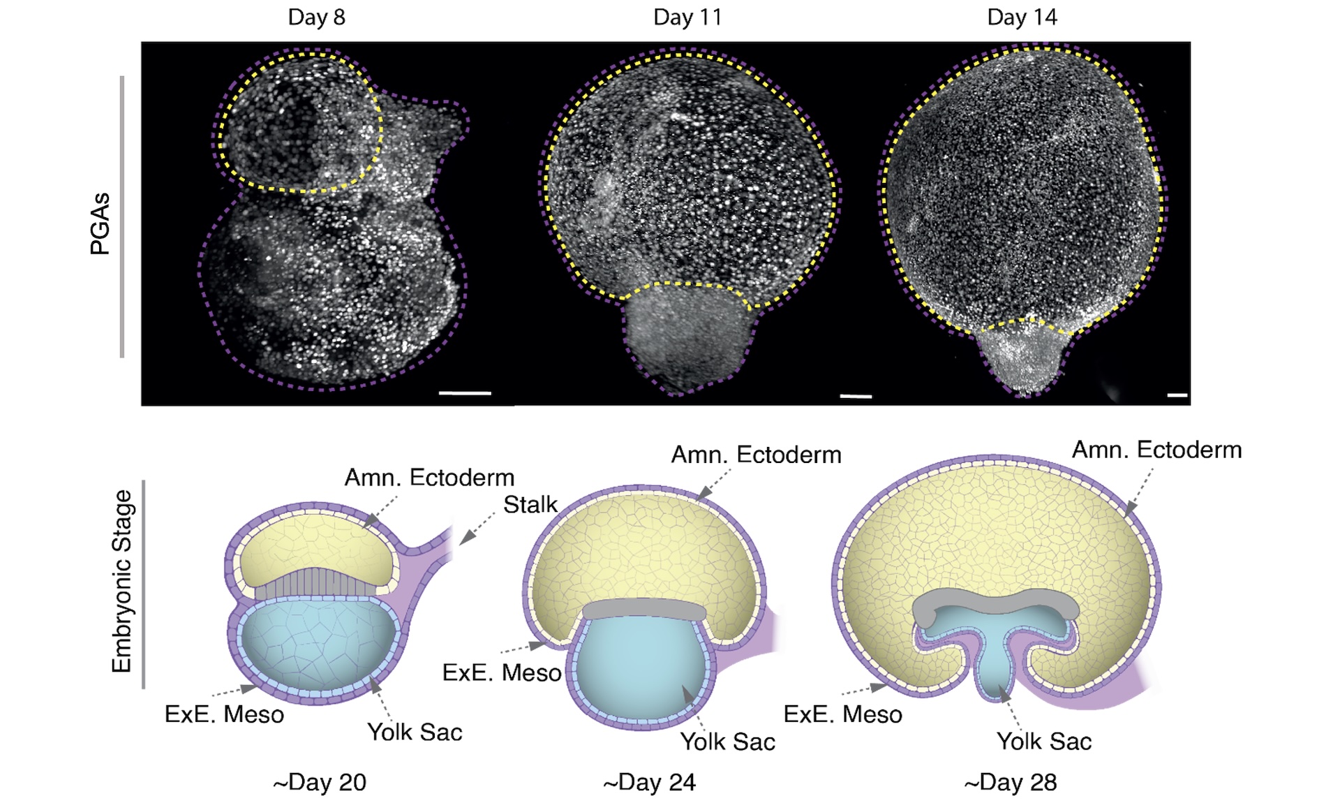
This diagram shows how the PGAs develop through time, largely through self-assembly. (Image credit: Gharibi, B. (2025). Cell.)
The Medical Applications
Amniotic tissues are already used in clinical settings, particularly in corneal repair, wound healing, as well as burn treatment, due to their antimicrobial & anti-inflammatory properties. But, sourcing Amniotic tissue generally depends on donations achieved from elective cesarean deliveries, which could be hard to scale and inconsistent.
On the other hand, Lab-Grown Amniotic Sac or PGAs provide a laboratory-grown, standardized, as well as renewable alternative that could rule out many of the logistical concerns. Moreover, the potential to develop these structures from iPSCs (induced Pluripotent Stem Cells), which are made by reprogramming adult cells, further raises the possibility of generating personalized tissues using a patient’s cells.
An Assistant Professor of Biomedical and Chemical Engineering at Syracuse University, Dr. Yi Zheng, stated that while further testing is essential, PGAs might have the potential to become a reliable source of clinically useful Lab-Grown Amniotic Sac, with numerous applications in Personalized Medicine and Regenerative Therapies.
Impact for Developmental Disorders
The capability to study as well as grow Amniotic Sac models in the laboratory can aid in understanding the causes of certain congenital disorders. Some development complications, including Congenital disabilities, are thought to be linked to abnormalities in fluid composition or Amniotic Sac formation during early pregnancy. By observing how PGAs function as well as develop over time, scientists may uncover the root causes of these early disruptions as well as create strategies for intervention or prevention.
This advanced Research opens new horizons into a previously inaccessible early phase of human pregnancy. It also offers a novel platform for both Therapeutic discovery as well as fundamental Research. The PGAs provide scientists a powerful model to ask as well as answer questions that were once impossible to inspect or investigate.
In her concluding statements, Dr. Santos said, “I’m extremely excited about the potential of these little structures.”
From revealing how life begins to transforming how we treat diseases and injury, these self-organizing and Stem cell-derived sacs represent a profound advancement. These are smaller in size, but have the potential to revolutionize the Life Sciences sector to a greater level.
To understand the full Experimental innovation and Scientific details, you are encouraged to explore the original Research by Dr. Silvia Santos et al. at the Francis Crick Institute, published in the journal “Cell.”

