


CSIR UNIT 1G Protein Conformation Ramachandran Plot Notes Download + Effective Study Method
If you’re preparing for the CSIR NET Life Science Exam, you know how important it is to master the conformation of proteins. This topic is covered in Unit 1G of the exam and includes concepts like the Ramachandran plot, secondary structure, domains, motifs, and folds. In this article, we’ll provide you with tips on how to study and remember these important concepts in preparation for the exam.
The study of proteins is an essential part of the life sciences, as proteins are the building blocks of life. Proteins are complex molecules that are made up of long chains of amino acids, and they fold into intricate 3D structures that determine their function. Understanding the conformation of proteins is crucial for many areas of research, including drug discovery, protein engineering, and molecular biology.
Today we will discuss how to study the conformation of proteins for the upcoming CSIR NET Life Science Exam and how to remember, practice, and recall important concepts and cycles. We will also elaborate on how to study the Ramachandran plot, secondary structure, domains, motifs, and folds, specifically with examples.
Proteins – CSIR NET Life Science
Proteins are folded into a specific 3D structure, which is known as its conformation. The conformation of a protein is determined by the sequence of amino acids that make up the protein. The folding process is driven by the interactions between the amino acids and the surrounding environment. These interactions include hydrogen bonds, van der Waals forces, and electrostatic interactions.
CSIR NET UNIT 1G Conformation of Proteins – Ramachandran Plot Notes
To study the conformation of proteins – CSIR NET UNIT 1G, we need to understand the different levels of protein structure. Proteins have four levels of structure: primary, secondary, tertiary, and quaternary. The primary structure of a protein is its sequence of amino acids. The secondary structure refers to the regular, repeating patterns of folding that occur within the protein chain. The tertiary structure refers to the overall 3D shape of the protein, which is determined by the interactions between the amino acids. The quaternary structure refers to the arrangement of multiple protein subunits into a larger protein complex.
How To Simplify & Study Ramachandran plot?
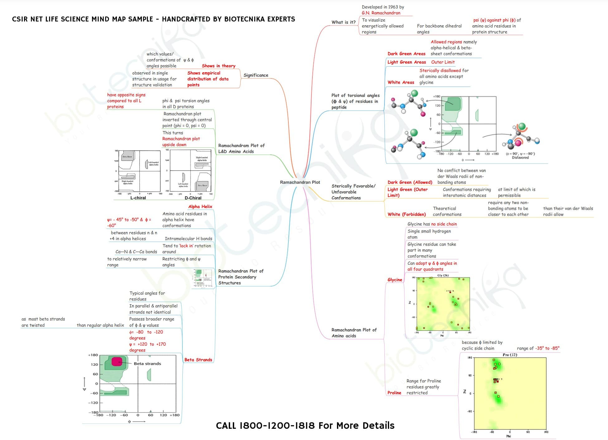
One of the most important tools for studying the conformation of proteins is the Ramachandran plot. The Ramachandran plot is a graphical representation of the possible rotation angles for a protein’s backbone atoms. It determines the allowed and disallowed regions of the protein’s conformation. The allowed regions correspond to stable and energetically favorable conformations, while the disallowed regions correspond to unstable and energetically unfavorable conformations.
Fast Track Revision Batch For CSIR NET June 2023 Exam
While studying the Ramachandran plot, it is important to understand the different types of secondary structures. The two most common types of secondary structures are alpha helices and beta sheets. Alpha helices are formed by a spiral of amino acids, while beta sheets are formed by a series of hydrogen bonds between neighboring strands of amino acids. The Ramachandran plot can be used to identify the regions of the protein that are likely to form alpha helices or beta sheets.
CSIR NET UNIT 1G Conformation of Proteins – Ramachandran Plot Notes
Another important aspect of studying the conformation of proteins is understanding the different domains, motifs, and folds. A protein domain is a distinct structural and functional unit within a protein. Protein motifs are short, conserved sequences within a protein that are important for its function. Protein folds refer to the overall 3D structure of a protein.
Quick Notes on Ramachandran Plot:
- The Ramachandran plot is a graph that shows the allowed regions of sterically allowed torsion angles (phi and psi) for amino acids in a protein. The plot was named after its creator, G.N. Ramachandran, who first described it in a paper published in 1963.
- The x-axis of the plot represents the phi angle, which is the angle of rotation about the N-Cα bond.
- The y-axis represents the psi angle, which is the angle of rotation about the Cα-C bond. Each point on the plot represents a single amino acid residue, and the color of the point indicates the frequency of occurrence of that residue in the allowed regions of the plot.
- The Ramachandran plot is useful for analyzing the conformation of proteins because it shows which combinations of phi and psi angles are allowed and which are not. For example, the plot shows that there are certain combinations of phi and psi angles that are not allowed because they result in steric clashes between the atoms in the protein backbone. These regions are known as the forbidden regions of the plot.
- The plot also shows that there are regions of the plot where the backbone can adopt certain secondary structures, such as alpha helices and beta sheets. For example, the alpha helix is characterized by a phi angle of -60 degrees and a psi angle of -45 degrees, which fall within the allowed region of the plot for alpha helices. Similarly, beta sheets are characterized by a phi angle of -120 degrees and a psi angle of 120 degrees, which fall within the allowed region of the plot for beta sheets.
- One of the challenges of using the Ramachandran plot is that it can be difficult to interpret. For example, the plot shows that there are many allowed regions for each amino acid residue, which can make it difficult to determine the precise conformation of a protein based on the plot alone. Additionally, the plot does not take into account the effects of side chain interactions on protein conformation.
- Despite these challenges, the Ramachandran plot remains an important tool for analyzing protein conformation. It can be used to identify potential errors in protein structures, to predict the stability of protein structures, and to design new proteins with desired properties.
CSIR NET UNIT 1G Conformation of Proteins – Ramachandran Plot Notes
To study protein domains, motifs, and folds, it is important to understand the different types of protein structures. There are four major types of protein structures: all alpha, all beta, alpha/beta, and alpha+beta. All alpha structures are dominated by alpha helices, all beta structures are dominated by beta sheets, alpha/beta structures have both alpha helices and beta sheets, and alpha+beta structures have separate regions of alpha helices and beta sheets.
It is important to use active learning techniques to remember, practice, and recall important concepts and cycles related to the conformation of proteins. Active learning involves engaging with the material in a way that promotes retention and recall.
CSIR UNIT 1G Protein Conformation Notes
Some effective active learning techniques For CSIR NET Life Science Exam Concepts include:
- Practice problems: Solving practice problems related to the conformation of proteins can help reinforce key concepts and cycles.
- Flashcards: Flashcards are a great way to memorize important terms and definitions related to the conformation of proteins. For example, you can create flashcards that include the definitions of protein domains, motifs, and folds, or flashcards that include the different types of secondary structure.
- Concept maps: Concept maps are a visual way to organize information and show the relationships between different concepts. You can create concept maps to help you understand the different levels of protein structure and how they are related to each other.
- Mnemonics: Mnemonics are memory aids that use patterns or associations to help you remember information. For example, you can use the acronym HAT BAT to remember that alpha helices are formed by a spiral of amino acids, while beta sheets are formed by a series of hydrogen bonds between neighbouring strands of amino acids.
- Practice exams: Taking practice exams can help you identify areas where you need to focus your study efforts. Many CSIR NET Life Science Exam study materials include practice exams that you can use to test your knowledge of the conformation of proteins.
In addition to these active learning techniques, staying organized and creating a study schedule is important. Make a list of the key concepts and cycles related to the conformation of proteins you need to study, and schedule time each day to review and practice them. This will help you stay on track and ensure that you are making progress toward your goals.
Finally, it is important to seek additional resources and support if needed. There are many resources available for studying the conformation of proteins, including textbooks, online courses, and study groups. If you are struggling with a particular concept or cycle, don’t be afraid to reach out to a teacher, tutor, or mentor for help. Biotecnika is always there for your support.
Join us on Telegram & WhatsApp groups for the latest CSIR NET updates, notes, flowcharts, study tools, test series, and more.
Check out the Koncept Series By Biotecnika
- KONCEPT Table For CSIR NET Life Science Exam
- Konceptika Lite – 100 Flowcharts on 100 Important topics of CSIR NET Exam
- CSIR NET Koncept Wheel Book
- Koncept Cloud – Latest Study Tool Revolutionizing CSIR NET Revisions
- Konceptika – The Flowchart Book
In conclusion, studying the conformation of proteins is an essential part of the CSIR NET Life Science Exam. Understanding the different levels of protein structure, including the Ramachandran plot, secondary structure, domains, motifs, and folds, is crucial for success on the exam. To remember, practice, and recall important concepts and cycles related to the conformation of proteins, it is important to use active learning techniques, stay organized, create a study schedule, and seek out additional resources and support if needed. With dedication and hard work, anyone can master the conformation of proteins and excel on the CSIR NET Life Science Exam.
CSIR NET UNIT 1G Conformation of Proteins – Ramachandran Plot Notes
CSIR UNIT 1G Protein Conformation Ramachandran Plot Notes PDF Download


 2026 Notification Released – Check Eligibility, Syllabus & Apply Online")


