Detailed Images Of Living Bacteria Using High Powered Microscopes
There is always more to understand and explore. This is true if you zoom in on microscopic organisms or if you zoom out to the depths of the Universe. In the field of science, you discover new questions when you find answers for many others.
And hence, scientists have used their advanced high-powered microscopes to observe the bacteria’s protective skin and its organization, disclosing new details about the bacterial skin.
Gram-negative bacteria such as Escherichia coli have external membranes to safeguard them from the commotion of bacterial life and hold their internals in the proper place. These protective layers have an excellent set of tools, including toxin and external-membrane proteins such as lipopolysaccharides present on the surface.
However, once you are familiar with this, you question more- what is the bacteria’s studs to membrane ratio? How it all fits together? This is where the new study comes into the picture.
Senior author and Biophysicist from the University College London, Bart Hoogenboom explained that the outer membrane is a crucial factor that makes pathogenic bacteria resistant to medications and is a strong barrier against antibiotics. Nonetheless, it is still obscure about the arrangement of this membrane, which is why they selected this area to explore in depth.
By examining live bacteria from their molecular to the cellular level, they can observe the formation of a network covering the whole bacterial surface by the membrane proteins, leaving tiny spaces for patches that have no protein.
The scientists employed an atomic force microscope for this study. AFM (Atomic Force Microscopy) consistently pokes the membrane’s surface to ascertain its shape. It is similar to reading braille, however with lasers rather than fingertips.
The group then produced a great microscopic image of the bacterial membrane- which they define as the sharpest images of a living bacteria ever produced- demonstrating the density of the proteins present throughout the surface of the outer membrane.
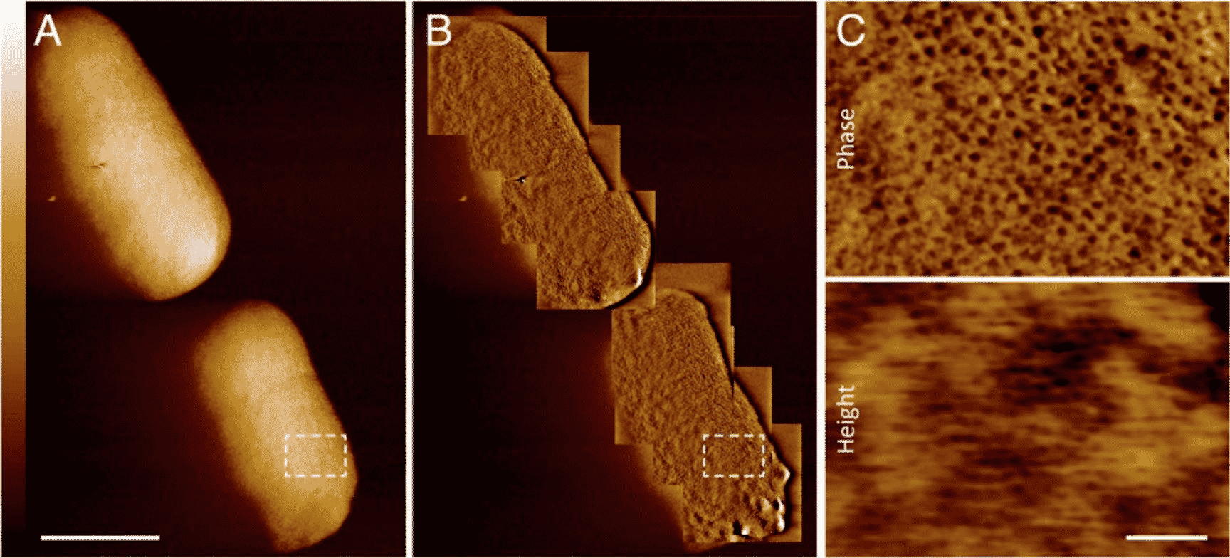
As you can observe in the sharpest image of E.coli above, there are many small holes throughout the surface. These are known are porins, beta-barrel proteins that form tunnels via the bacterial membrane enabling the entry of certain molecules. The small parts of the smooth surface are the expandable lipopolysaccharides which stretch during the cell division.
The research team mentioned in the article that the outer membrane is a mix of outer-membrane protein-rich and phase-segregated lipopolysaccharide-rich regions. The maintenance of these is important for the membrane integrity and therefore for the gram-negative bacteria’s lifestyle.
Obviously, researchers do not observe E.coli using advanced microscopes simply for fun. There are certain reasons why they require this degree of detailed apprehension.
Georgina Benn, Biochemist from UCL and the main author of the study elucidated that the textbook image of bacteria’s outer membrane indicates the disordered distribution of proteins across the membrane which is well-integrated with other membrane’s building blocks. Their images, on the other hand, contradict this depiction. It shows that is lipid patches are separated from protein-rich networks similar to the segregation of oil in water, which for a few cases forms chinks in the bacteria’s armour.
This new method of observing the outer membrane infers that now people can begin examining how and if this particular arrangement is important for the antibiotic resistance, integrity, and function of the membrane.
The group will be studying how this new information can be employed to outwit antimicrobial resistance in gram-negative bacteria like E.coli.
The new findings have been presented in the journal, PNAS.
DOI: 10.1073/pnas.2112237118
Detailed Images Of Living Bacteria, Sharpest Image Of E.coli
Further Read: Pune Based Researchers Detects 108 Mutations Of SARS-Cov2 In Wastewater Samples