


Imaging Embryo Orientation – Witnessing The Emergence Of The First Body Plan
Eggs that initially appear like circular blobs undergo a transformation into the particular species upon fertilization by orienting in specific directions – left to right, back to belly, or head to tail. Scientists have speculated these directions of body orientation that hadn’t been seen till recently. The scientists at MBL (Marine Biological Laboratory) have successfully performed the imaging of this cellular rearrangement beginnings, and this study has helped answer some fundamental questions in biology.
The very origin of an animal’s body axis is developmental biology’s most mysterious and interesting phenomenon, as MBL scientist Tomomi Tani says. At the National Institute of Advanced Industrial Science and Technology in Japan, Tani performed the research while being at Eugene Bell Center. The offspring’s body orientation is influenced by both parents, as the study by Hirokazu Ishii and Tani, which was published in Molecular Biology of the Cell, said. In the sea squirts that were the study organisms here in imaging embryo orientation, the back-belly axis was generated from the mother while the father contributed the head-tail axis.
Tani explained that the body plans for an animal embryo’s development required cues from both the mother and father. This research which seeks to understand solutions for vital problems in developmental biology could aid in developing fields as diverse as agriculture and medicine.
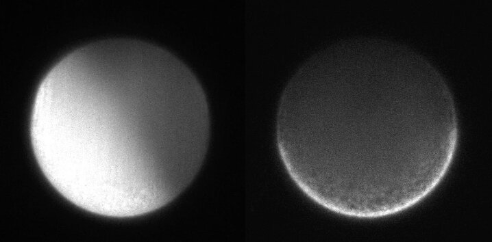
Left: Fertilized Ciona egg showing calcium waves transmission. (Selective Plane Illumination Microscopy [SPIM] movie showing calcium ion indicator fluorescence, Rhod-dextran). Right: Same egg’s autofluorescence image showing cytoplasmic motion. Each frame was taken every 2s. Replay speed, 10 frames/s. Credit: Hiro Ishii and Tomomi Tani.
The widespread concept of how the body axis is established is based on cell movement and contraction governed by the egg’s actin filaments. These facilitate cytoplasmic rearrangement following fertilization. Since the area of this rapid activity is extremely minute, visual observation has been a challenge until recently imaging embryo orientation.
Rudolf Oudenburg, senior scientist at MBL, and Shalin Mehta (now working in Chan Zuckerberg Biohub), and Tani developed a technique called fluorescence polarization microscope a few years ago, which is what was used to overcome visualization problems by Tani and Ishii. This method, familiar to the MBL scientists, gives us the ability to observe and image processes at nanometric distances. In fact, it seems to be MBL’s tradition to use polarized light to observe molecular order dynamics beginning in the 1950s Shinya Inoué’s pioneering research on live cells.
Thus, they used polarized light waves that oscillate completely or partially in a single direction – clockwise/anticlockwise, left/right, up/down, etc. This is why polarized light in one orientation can pass through a filter, but not when rotated. Fluorescent probe molecules were attached to the actin in the sea squirts’ (Ciona) eggs by the 2 scientists for imaging embryo orientation. Tani explained that the rigidity of the probe-actin link enabled microscopic detection of orientation using the polarized light.
Left: Ciona egg before and after fertilization (Movie of Total Internal Reflection Fluorescence Microscopy [TIRFM] images; F-actin probe is AF488-phalloidin). Right: F-actin alignments’ transient changes before fertilization to egg’s first division. Positions of AF488-phalloidin particles bound to F-actin are represented as yellow dots, and F-actin orientation is represented as yellow bars. Frame intervals, 10s. Replay speed, 15 frames/s. Credit: Hiro Ishii and Tomomi TaniThe researchers could thus observe if the actin lay jumbled or was oriented in a single direction. An unfertilized egg showed random actin orientation, while a calcium ion wave transmitted through the egg after fertilization displayed a particular actin arrangement. The future back/belly axis was seen to be at a right angle to the actin filaments, followed b the movement of the cytoplasm.
Other investigations are being held to follow up to identify and comprehend the developing embryo’s drive to shape its form, morphology, and structure. Tani discussed future goals of finding the morphological organization in animals governed by the cytoskeleton’s molecular order.
Imaging embryo orientation


