


Imaging Quadruple Helix DNA In Living Human Cells For The First Time
Scientists have tracked the formation of four-stranded DNA in living human cells to see how it works and study its role in cancer.
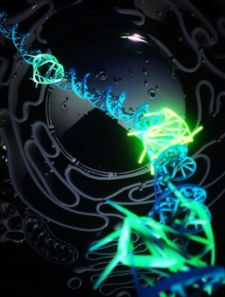
The double helix structure of DNA was discovered in 1953. DNA usually form this structure by wounding two strands around each other. Several other structures were found in test tubes, but that doesn’t imply they are found in living cells also.
Cells were detected previously with DNA G-quadruplexes (G4s), which are the quadruple helix structures.
Quadruple helix structures, called DNA G-quadruplexes (G4s), have previously been detected in cells. However, the technique involved using high concentrations of chemical probes to visualize G4 formation or killing the cells. Due to this, tracking the presence of such quadruple helix structures in living cells under normal conditions was difficult.
A team of scientists from Imperial College London, the University of Cambridge, and Leeds University have developed a fluorescent marker that is able to attach to G4s in living human cells. The marker, for the first time, enables scientists to visualize how the structure forms and what role it plays in cells.
Nature Chemistry published the study today.
For the first time, researchers are able to prove the existence of quadruple helix DNA in human cells as a stable structure generated by normal cellular processes, said Dr. Marco Di Antonio, one of the lead researchers and now leading a research team in the Department of Chemistry at Imperial. This discovery forces scientists to rethink the structure of DNA. This new area of fundamental biology could open up new insights in therapy and diagnosis of medical conditions like cancer.
Now the scientists can ask directly what the real biological role of G4s is as they can track them in real-time in cells. G4s are seen more prevalent in cancer cells, and now scientists can develop new cancer therapies targeting G4s.
The researchers think that G4s could be forming to temporarily hold the DNA open to facilitate transcription like processes so that the DNA instructions can be read and proteins can be made. This could be a way of activating a part of the genetic code or a form of gene expression.
G4s are detected in large numbers in cancer cells and are often associated with genes involved in cancer. Now the team can track the role of G4s within specific genes and cancer cells as they can image a single G4 at a time. Drugs that interrupt the formation of cancer could be developed with the knowledge of new targets.
Imaging Quadruple Helix DNA
Previously, scientists were using high concentrations of molecules and antibodies that could find and attach to G4s. But this used to disrupt the DNA, forcing them to form G4s rather than detecting natural G4s.
Dr. Aleks Ponjavic at the University of Leeds led the research and developed the new method of visualizing fluorescent markers with microscopy. Scientists require probes to see molecules within cells, but sometimes these probes can disrupt the molecules inside. But now they can see probes at 1000-fold lower concentrations than previously used with the help of single-molecule microscopy. In this new technique, the probe binds to the molecule only for milliseconds, without affecting its stability. This allows them to study the behavior of G4s in their natural environment, without any external influence.
The researchers used a very bright fluorophore in small amounts that can bind to G4s very easily. The technique cannot visualize every G4 in the cell but instead identifies single G4s. By imaging quadruple DNA helix inside cells, the fundamental biological role of G4s can be studied using this technique, without disturbing their stability and overall prevalence in the cell.
The G4s form and dissipate very quickly. This suggests that they perform a certain function and could be toxic to normal cell functioning if they lasted long.


