


Images of Coronavirus Released by CDC – From Very First American COVID-19 Patient
All of us have seen pictures of the SARS-CoV2 coronavirus, yet until now, the majority of these images have been artist’s diagrams impacts and impressions. CDC has released coronavirus images from samples of the first American case of COVID-19. The picture gives incredible details of the virus.
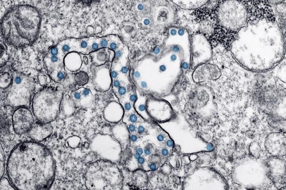
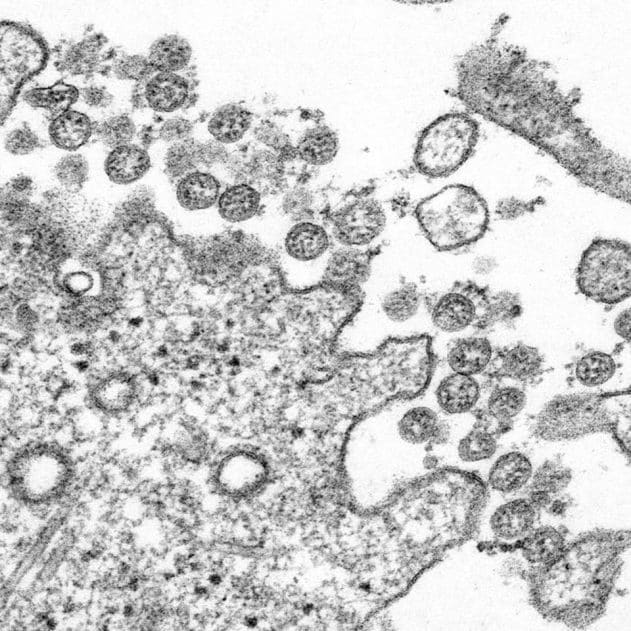
The image of the SARS-CoV2 virus looks like tiny pepperoni pizzas (blue in color in the image above), and these pictures are from the CDC Public Health Image Library. However, these images are hard as well as labor-intensive to generate, and for lots of people with COVID-19, photos like this will never be made.
Bryan William Jones, Ph.D., Associate professor, Ophthalmology and Neuroscience, University of Utah, who has been using electron microscopy for over two decades, said: “If you intend to see and also be able to identify viruses, much less determine any detail on them which aids you to classify them, you have to use an electron microscope”.
Using a microscope to identify the type of pathogen is the conventional way to diagnose patients with some contagious diseases. However, viruses are too small to see through the standard microscope, so this method is utilized primarily for infections, including fungal and bacterial.
Jones said, “There are lots of viruses with different proportions. The viruses with the largest size are about 500 nm, which implies that light microscope, you might just see them as dots. The smallest viruses are around 20 nm – you might never ever see them in a light microscope”.
Presently, the COVID-19 coronavirus is detected using a PCR test, which looks for traces of the viral genetic material in the samples. The CDC’s new images of coronavirus are taken using microscopes that utilize electrons and magnets to focus and produce pictures, as opposed to the traditional microscopes that use light and glass lenses.
Now the question is – So why can’t an electron microscope be used to test coronavirus? The foremost reason is that these microscopes are incredibly costly to purchase – around $1 million for a brand-new, top of the model, so not budget-friendly for many diagnostic laboratories. Secondly, they are usually huge compared to the majority of standard laboratory microscope nad also need highly-trained users. Additionally, the scientists may not be able to distinguish the SARS-CoV2 coronavirus from other coronaviruses causing common colds.
So, needless to say, the method is not practical, inaccurate, and unnecessary to use for diagnosis across the board. But in research settings, electron microscopes can aid scientists to understand the viruses better.
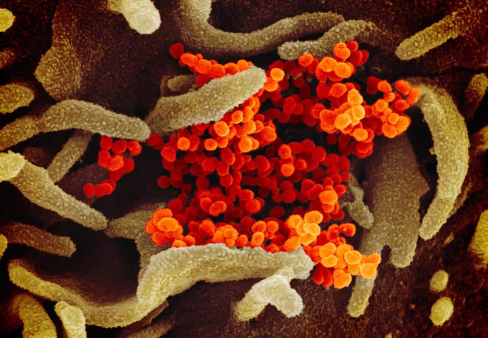
Jones added, “The shape or morphology of the virus is one more necessary method of categorizing types of virus, and this is where electron microscopy is essential. The SEM images let us see the 3D structure of the COVID-19 viruses, which include the nucleocapsid protein (N-protein), and a small fuzz present on the outside of the coat – the spike protein.”
NIAID-RML
Electron microscopes are of two types – TEM (Transmission electron microscope) and SEM (Scanning electron microscope), TEM produces a flat image, and SEM provides 3D-like photos. The pictures from all the electron microscopes are black and white, unlike light microscopes. The below picture has not been artificially colored, and colors are added later on to draw attention to particular elements of the image.
Jones said, “The blue dots in the TEM COVID-19 image (title picture above) and also the orange blobs in the SEM COVID-19 image (listed below, from the NIH), as an example, have been artificially colored to more easily reveal them to viewers who may not be experienced virologists who might understand specifically what to find.”
Author: Sruthi S


