


Ultrasound Spots Cancer Early
A new ultrasound technique is being regarded as the most significant development in technology for more than 60 years.
The Scientists at Heriot-Watt University in Edinburgh have developed this New ultrasound technique that is ten times better than current scans.
Researchers believe that its ability to precisely detect tumors could one day replace biopsies in investigating suspected cancer cases.
Ultrasound Spots Cancer Early- The History
In 1958, Prof Ian Donald of Glasgow University discovered the use of ultrasound in medical sciences to reveal how babies were developing in their mothers’ wombs. Ultrasound waves are not in the hearing range of human beings. Prof Ian Donald used the pulsed waves to produce images which were breakthrough research in the field of medical science.
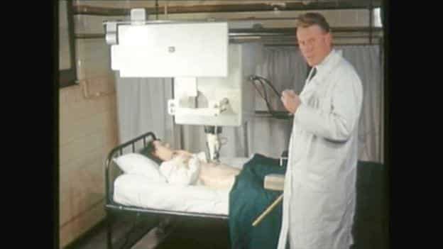
Women at Glasgow Royal Maternity Hospital were the first to benefit from a technique which is now common worldwide.
Though ultrasound has its limitations even after being discovered decades ago. The image resolution is low, and sometimes they are so indistinct it can be difficult to tell if the unborn child is a girl or a boy.
Ultrasound Spots Cancer Early- The New Technique
Dr. Vassilis Sboros says that this is the first technology that can claim to have a near-microscopic quality. He adds that in the future, this technique would be widely used.
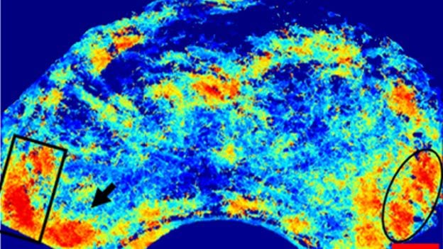
Alongside a familiar-looking greyscale scan, there is a new, far sharper color image produced by the new technique. The image is said to be 10X higher resolution.
Dr. Vassilis Sboros describes the process as like being able to look inside the body with the help of a microscope
Ultrasound Spots Cancer Early- The Principle
Scientists have used the technique used in physics, statistics, and bubbles.
Doctors have long used microbubbles to increase the contrast of ultrasound images during clinical studies. These are typically tiny capsules of hydrocarbon gas in a lipid shell, each bubble a fraction of a millimeter across. Clouds of them are injected into a patient’s bloodstream to diagnose liver and other diseases.
The team of scientists first used physics to observe the behavior of individual microbubbles. These microbubbles are the size of red blood cells.
Once the team had established the physics of microbubbles, Dr. Weiping Lu used statistics and computing power to reveal what ultrasound scans had not been able to show before.
Dr. Lu says it helps to think of each microbubble as a car in traffic. He used artificial intelligence to create a robust algorithm that can track each one and reveal the busiest routes.
He imagined the car as microbubbles, and the road network is a blood vessel. They monitored the speed of these ‘cars.’ The scientists also measured the ‘roads’ that is the blood vessels.
Just as tracking individual cars would build a road map, this signal processing produces an image of blood vessels by watching where microbubbles go.
How do they detect a possible tumor formation?
Dr. Lu highlights that if they go somewhere unexpected, it could be a sign of cancer. It has the potential to be detected much earlier than before. These are super-resolution images showing details far beyond the limitations of the conventional scanner.
In normal conditions, scanning a patient’s abdomen would show details as minute as a millimeter. The new images are already ten times better, and the team expects to be able to refine the process to see more minor features yet. Trials on human patients are scheduled to begin before the end of 2019 at the Western General Hospital in Edinburgh.
Ultrasound Spots Cancer Early- The Applications
With the help of this new technique, doctors can find patients who need urgent treatment.
Prostate Tumor patients are being tested first because a patient’s gland is typically removed entirely during surgery. This means the accuracy of the new technique can be compared with the real-time scenario.
Most essential diseases change the body’s blood flow, which means this technique could be used across medicine and may not be limited only to cancer detection.
The results have been published in the Journal of Investigative Radiology
The research was funded by a Science and Technology Facilities Council project. The project was called ‘Imaging The Stars From Within.’


