Super-Resolution Microscope
A microscope is an instrument that produces enlarged images of tiny objects. This allows the observer an exceedingly close view of minute structures at a scale convenient for examination and analysis.
Generally, the microscope is slow in processing images involving extensive data and high-resolution images.
Researchers have developed a way to speed up the process by creating a method which can record data at this microscopic scale in real-time.
The most significant advantage of a faster microscope- The Super-Resolution Microscope is that it can be used to observe the movements of small objects like different structures of cells.
Professor Dr. Thomas Huser, head of the Biomolecular Physics at Bielefeld University, said that the speed and accuracy of this microscope make it highly useful for applications in biology or medicine. Dr. Huser is the first author of the paper.
He added that the major problem so far was that microscopes offering high resolution could not display information at the corresponding speed.
Principle Behind The Super-Resolution Microscope
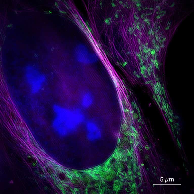
The super-resolution microscope works by irradiating objects with laser light, so the molecules in the sample are excited and glow at different wavelengths. The microscope then picks up this re-emitted light. Due to this reason, it provides a higher resolution image than other methods as well as a quick result.
Andreas Markwirth, the lead author of the study, said that the team of researchers able to produce about 60 frames per second. This is a higher frame rate than the cinema films. The super-resolution microscope allows real-time recording; therefore, the time between measurement and image is less than 250 milliseconds.
This real-time recording method used by the super-resolution microscope can be used to examine different biological elements, including the less-studied phenomenon such as movements of mitochondria. It can also be used to observe the way that viral particles move through a cell.
Dr. Huser highlighted that the newly discovered method by his team has both high-resolution and fast imaging process. This enables biologists to explore such structures.
Dr. Huser and team are planning to use the new technology for various kinds of research in the area of Cell Biology.