


")
Stem cell for human development- A New System Developed By Scientists
A new method for making stem cell colonies that mimic early human development could help investigate critical questions in maternal and child health.
Scientists at the University of Michigan developed this technique. It imitates stages in embryo development that occurs after implantation. This is when the amniotic sac begins to form, and when the stem cells that would go on to become the fetus take their first steps toward the organization into the body.
The embryo-like or “embryoid” structures don’t have the potential to develop beyond small colonies of cells.
The system can reliably produce thousands of embryo-like structures required to determine whether a drug is safe for a pregnant woman to take during pregnancy. This system will answer longstanding questions such as- What chemicals pose risks to developing embryos, and what causes specific congenital disabilities and multiple miscarriages?
Stem cell for human development- An Overview
Jianping Fu, an associate professor of mechanical engineering, led a team of engineers and biologists. They arranged the stem cells into organizing and behaving in ways that resemble certain aspects of embryonic development.
The Scientists at the University of Michigan developed these three models:
- The epiblast, a colony of stem cells that comprise most of the cells that would go on to form the fetus.
- The beginnings of the amniotic sac and the posterior, or rear end of the epiblast after it has been through the very first steps of differentiation. In an actual embryo, these posterior cells go on to become the lower portion of the fetus.
- The beginnings of the amniotic sac and the anterior, or upper end of the epiblast. This stage is marked merely by the absence of posterior cells. In an actual embryo, these cells would go on to form the upper portion of the fetus, including the head and mid-section.
Fu added that stem cell structures that mimic embryos could help fill critical gaps in knowledge about early human development. This research will give scientists a window into the pivotal but barely observable period between two and four weeks after conception. Study confirms that this is a time when many miscarriages happen, and severe congenital disabilities can form. Researchers have even begun to find connections between late-onset diseases and early development. Researchers need to understand this process deeply to design a preventive measure.
Fu says the new system is ready to screen medications for safety during early pregnancy. He added that the lack of knowledge about how drugs affect embryo development is a severe public health problem. In his opinion, this research work provides a controllable and scalable experimental platform to ask important questions related to human development and reproduction
If the team at the University of Michigan can continue this research, it could also give insights into the causes of some congenital disabilities.
The system works with reprogrammed adult cells as well as embryonic stem cells; it may also be able to shed light on some serious issues such as infertility. At present, around 30% of couples who seek fertility treatment don’t receive an explanation for why they haven’t conceived.
Fu highlighted that human development is very different from other mammals. Different signaling pathways are involved. So this is the only way to accurately study human development without using intact human embryos- that will include ethical issues.
Stem cell for human development- How Does this System Work?
A microfluidic system that allows stem cells to self-organize
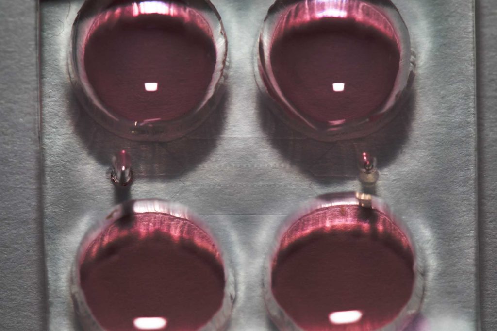
To generate the models, the team of scientists grew stem cells in a scalable microfluidic system of three channels. The central channel consisted of a gel that mimicked the wall of the uterus, and a trough flanked it for feeding in stem cells and another for chemical signals that guided the development of the cells.
The stem cells loaded into the microfluidic device was first grown into colonies that naturally became hollow balls. The balls were burrowed into the gel, similar to the way that an early embryo implants in the wall of the uterus. This stage comprises of the first model.
To generate the second model, the researchers loaded into the microfluidic device chemical signals that triggered the stem cells on one side of the ball to turn into amniotic sac cells.
To generate the third model, the researchers added to the microfluidic device molecules that block the undifferentiated epiblast cells from evolving into rear epiblast cells. This mimics a signal that arises in real embryos.
Fu and his team hope to use this new system for clinical applications and design powerful diagnosing techniques for developmental defects.


