MRI to detect aging by providing biological readouts
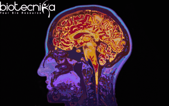
In recent research published in Nature Communications, a group of scientists have successfully transformed an MRI from a diagnostic camera into a device that can record changes in the biological makeup of brain tissue. This paves the way for doctors to understand if the patient is displaying signs of aging or developing any neurodegenerative disease like Alzheimer’s or Parkinson’s.
Dr. Mezer from the Hebrew University of Jerusalem said that the quantitative MRI model provides molecular information about the brain tissue rather than just the images. This allows the doctors to study and compare the brain scans of the patients taken over a period of time and differentiate the healthy and diseased brain tissue without the requirement of any expensive and invasive procedures like biopsies.
The familiar signs of aging like the gray coloration of hair stooped spine, occasional memory loss can be observed externally. But we do not know if the patient’s brain is undergoing healthy aging or developing disorders. This can be understood on the biological level. The changes in the brain create “Biological footprints” by changing the lipid and protein content in the tissues.
The new MRI technique developed promises
to provide biological readouts of brain tissue where the activities on the molecular level can be observed to detect aging, and a course of treatment can be designedShir Filo, a Ph.D. student part of this study, explained that A blood test gives the exact number of white blood cells which can be compared to the normal value to diagnose the illness. In the same way, changes observed in the composition of the human brain can help in differentiate normal aging from the brain composition of a patient with the onset of degenerative disorders.
Mezer also believes that the new MRI technique will also provide a crucial understanding of how our brains age. It was also observed that the different areas of the brain undergo the aging process differently. For example, some white-matter areas showed a decrease in brain tissue volume, whereas, in the gray-matter, tissue volume remains constant.
This technique will be a blessing for the patients and will pave the way to early and correct diagnosis, speed up the begin of treatment, ultimately maintaining and improving quality of life.


