


Cryo-EM in Biological Research -The ‘New Resolution Revolution’
The technique was once termed “blobology” because the images were so indistinct. But recently this groundbreaking technology is being used to create videos of the body’s inner-workings as well as to know what happens at near-atomic levels or in other words, this at a hitherto unseen level of detail. With this technology, the views inside the body are likely to accelerate the development of more effective drugs against dementia and infection.
Dr. Peter Rosenthal from the Francis Crick Institute in London said to BBC News that there is a growing buzz about the potential of the technology and this technology is the new hottest approach in biological research at the moment.
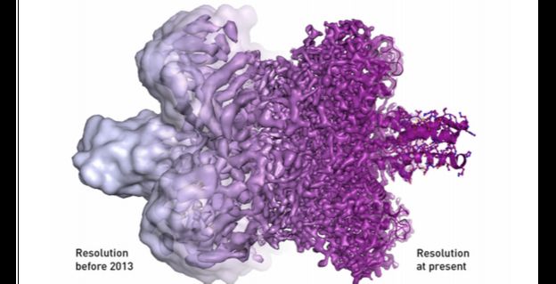
This technology is been described now as a ‘resolution revolution’. With this technology in biological research, there is an exponential growth in the number of new images as well as 3D maps being produced and researchers are clamoring to be trained how to do it.
This was not always so for the technique, called cryo-electron microscopy, cryo-EM for short. Cryo-EM in biological research was seen as a wacky alternative to all other techniques for decades. And this was because the resulting images from most of the techniques were little more than blobs. Thus came the rude description of “blobology” by its detractors.
But over time, researchers refined cryo-EM in biological research to the point that it earned a Nobel Prize for chemistry in 2017. But there was a perfect storm of improvements in camera technology as well as image processing by reduced cost and also increased computing power which had a major role in utterly transforming “blobology” into an “ultra-high-definition-3D-video-ology”.
The videos by Cryo-EM in biological research are showing scientists what is actually going on inside cells and this is paving the way for the development of better drugs for infectious disease. These include even those diseases that are challenging to treat like flu, HIV/AIDS as well as many of the diseases associated with dementia-like the Alzheimer’s and Parkinson’s diseases.
Dr. Rosenthal said that these videos from this technology are like seeing how all the different parts of an orchestra play to create a piece of music. Scientists will be able to learn how processes in cells work in great detail and also what goes wrong when something gets out of tune or the normal pattern and causes diseases.
Lesley Calder, a flu researcher at the Crick Institute, said that cryo-Electron Microscopy in biological research has burst on to the scene and is transforming her research studies. Cryo-EM in biological research is a huge step forward in being able to see what is going on within structures and where, before, cutting them into slices or could just see the outside of. Now one can see the whole object, she added.
This is not just the case with Lesly. Many Scientists using this technology, the funding available for it and the number of images and videos posted has all recently spiked.
According to Dr. Donald Benton, there is lots of exciting biology coming out every week from these high-resolution Cryo-EM microscopes in biological research. Researchers can now see the structure of the molecules in action within the cell.
Seeing the shapes and structure of biological molecules as well as cell at an atomic level are important because those are the cogs and wheels that make any living things work. They work inside and between cells. As we all know cells are the building blocks of human life. Many of the human body’s basic processes occur in cells. Cells are where raging wars against diseases are fought.
Traditionally, scientists were using a process called X-ray crystallography in biological research to see what goes on at cells or near-atomic levels. The process of X-ray Crystallography involved turning the sample into a crystal and beaming X-rays at it for visualization.
The X-rays then scatter when they bump into the sample’s molecules and scientists were able to reconstruct a highly accurate picture of the sample from the pattern of the scattered X-rays.
The X-ray crystallography technique has been incredibly successful. The technique produces images of the structure of biological molecules at atomic resolution. It has almost revolutionized one’s understanding of biological processes and this also led to hundreds of new medicines. The drawback of this technique is that the molecules are taken out of their natural environment. This technique gives a snapshot of its shape rather than an observation of what it does in the cell.
Cryo-EM in biological research -New Resolution Revolution- on the other hand, enables researchers to see the molecules in their natural state and even observation of what it does in the cell. The process of cryo-EM, as the name indicates, involves freezing biological molecules or viruses inside the cell and then taking thousands of pictures of these frozen materials from different angles and at different stages in the biological process using a Cryo-electron microscope. Scientists can then stitch these pictures together and create a video of living processes in action inside the cell!


