Flow Cytometry – Working Principle, Applications & Job Opportunities
Flow cytometry is an advanced analytical technique which has been used for the analysis of single particles or cells. Diverse physical characteristics like internal complexity or relative granularity, relative size and relative fluorescence intensity are measured using a beam of laser light and analyzed as the single particles or cells flow in the stream of fluid to the laser interface. Particles in the range of 0.2–150 micrometers are fit for the Flow cytometry. The particles should be properly suspended in the fluid stream such that they could be directed to the laser beam in single file. As the particles pass through the laser intercept, light is scattered and in presence of fluorochrome particle emits fluorescence light. These scattered and fluorescent lights are collected and converted to electrical signals employing an optical-to-electronic coupling system.
Flow cytometry has been extensively used for the analysis of cells. The earliest experiment was conducted in the 1950s which involved the measurement of cell volume using this technique for cells present in a rapidly flowing fluid stream. Since then many more modifications and advancement have been done to the technique to create the present-day flow cytometry. Today’s advanced flow cytometers have higher efficiency, precision and accuracy with the rate of detection as high as 10,000 cells/second and can detect many fluorescent parameters at a time. It allows a multi-parameter analysis of single cells simultaneously. Because of all these advantages flow cytometry is the most preferred tool for cell biologists. It is widely being used to scrutinize and characterize millions of cells within a short span of time with greater efficiency at a comparable cost to other existing microscopy techniques.
The popularity of this analytical tool can be estimated by its range of uses. It has been used for cell counting, cell sorting, measuring cell size and volume, characterizing different cell types in a mixed cell population, analyzing both cell surface and intracellular molecular expression and assessing the purity of isolated subpopulations of cells. Principally checks the fluorescence intensity generated by fluorescent-labeled probes associated with proteins, lipids, DNA or any other specific cellular molecules. In this artcile, we will throw some light on the Flow Cytometry – Working Principle, Applications & Job Opportunities which you can explore if you are skilled in Flow cytometry.
Flow Cytometry – Working Principle
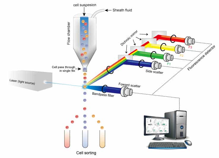
The basic working principle of Flow Cytometry is – this analytical technique requires fluorescent labeling of cells, followed by the passage of these cells suspended in a fluid sheet as a single file to a laser beam. The scattered and fluorescent wavelength are detected and analyzed or used for sorting of cells.
Sample preparation is one of the foremost steps. The preparation protocol differs based on the source, target, and purpose. In case some tissue is used as a source of cells then it should be first disintegrated into single cells, but when blood is source then no need of disintegration. Further, it is important to tag a fluorescent probe mostly an antibody which can detect cell-specific protein. Many cases commercially available antibodies are used. Sometimes unknown antibodies can be developed and used. But for both prior to flow cytometry microscopic evaluation of specific and non-specific staining or any abnormal staining is important. Once the staining is optimized using microscopy, it can be used for sample preparation in flow cytometry.
Next, an important step in the flow cytometric analysis of these sample cells. The three principal components of a flow cytometer are:
1. Fluidics,
2. Optics, and
3. Electronics.
1. Fluidics:
Fluidics, as the name suggests, is the fluid component of Flow cytometry. The sole purpose is to transfer cells/particles via this fluid stream to the laser interface. It is important to focus the cells or particles at the center of the beam and the cells or particles should be in single file to obtain optimum illumination. Fluidics comprises of two types of fluid – a sheath fluid which is mostly a saline buffer and the sample fluid which contains the cells or particles.
The sample is injected into the stream of fluid sheath forming the core of the stream which passes through the flow chamber known as flow cell or nozzle tip. The sampled core remains separate from the sheath fluid, but both remain coaxial in the flow chamber. Sheath fluid accelerates the sample by the principle of hydrodynamic focusing. Both the fluids have a pressure difference with the sample fluid being at higher pressure. The pressure of the sample fluid can be increased or decreased which can affect the measurement and resolution. Increasing the pressure widens the core sample stream, which leads to lowing of the resolution. This might be used for qualitative studies like immunotyping. Decreasing the pressure narrows the core stream, which increases the resolution. This is useful for studies where resolution is important like DNA analysis.
For proper measurement of data, it is very important to maintain the fluidics mainly pressure of both streams needs to be optimized, the choice of fluids such that it should not give any fluorescence, and the fluids should be of high purity devoid of air bubbles and debris.
2. Optics:
Optics of the flow cytometry is segregated as excitation optics and collection optics. Excitation optics comprises of laser source and lenses which create and focus the laser beam for optical measurement. Collection optics comprises of lenses to collect scattered or emitted by the particles or cell upon interaction with a laser. It also carries a system of mirrors and filters to direct specific wavelengths to detectors.
The beam of laser used for illumination is produced by different sources. Most commonly used is the argon laser which produces blue light. Some instruments use a helium-neon laser which generates red light, substituted with a mercury arc lamp. The light is passed through lenses for focusing and shaping to create an elliptical beam which has high intensity at the center. The beam is focused on the center of the flow chamber which has a quartz cuvette of about 200 µm inner diameter. The sample fluid passes through this as the laser strikes it.
Cells or particles interact with the laser beam and cause scatter and fluorescence. Scatter is of two types Forward scatter and side scatter. Forward scatter is mainly the diffracted light and is correlated to cell surface area and size. Side scatter is mainly refracted and reflected light and correlates to internal complexity or granularity of the cells. These could be used to distinguish between cells is population.
Fluorochromes like fluorescein isothiocyanate, phycoerythrin, peridinin chlorophyll protein, and allophycocyanin, show fluorescence upon absorption of light of specific wavelength. These fluorochromes emitting light of different wavelengths can be used to tag monoclonal antibodies, which can be used as a marker specific probe. Thus, from mixed population cells of different types can be distinguished and even sorted.
All these scattered or fluorescent lights are split into defined wavelengths and channeled by a set of filters and mirrors to the detectors which is either a photomultiplier (PMT) or photodiode (PD).
3. Electronics:
Electronics converts the optical measurement into electrical signals which could be further recorded and analyzed. The scattered and fluorescent wavelengths reach the photodetectors PMT or PD. PD is less sensitive to PMT, so generally used for detecting strong signals whereas PMT is more sensitive so used for weak signals. As the light of specific wavelength falls on the detector it generates electrons of equal proportion. This electrical current so generated is further amplified to create a voltage pulse. Amplification is of two types linear – 200 to 1000 fold and logarithmic- 2000 to 10000 fold. Till the particle is interacting with the laser beam, the pulse will be created reaching peak value when the particle is at the center of the beam and as it exits the beam voltage pulse comes down to baseline. The voltage signal could not be fed into the computer so first it is converted into a digital signal by an analog-to-digital converter (ADC). The digital signal is in inform of channel number which can range from 0-1023. This channel number can be transferred to the computer and displayed as data plot.
The collected data are stored in a prescribed format developed by the Society for Analytical Cytology in the computer and used for analysis. Saved data can be displayed in different formats like FCS, SSC, Fluorescence, etc. Analysis can be done employing histograms or dot blots. The data can be fragmented by gating or in the form of quadrants. The data analysis can further be coupled to cell sorting as well.
Applications of Flow Cytometry
Flow cytometry can be used for multiple purposes. Some of the potential applications are:
1) Estimation of cell number in a homogenous or heterogeneous population.
2) Identification of different cells in a heterogeneous population and their characterization.
3) Measurement of the level of protein expression in cell or nucleus
4) Monitoring, identifying and detection of post-translational modifications in protein like proteolytic cleavage, phosphorylation, glycosylation, acetylation, etc.
5) Detection and measurement of different RNA including mRNA, miRNA, etc.
6) Monitoring the health condition of cell-like viability to apoptosis
7) Monitoring the cell cycle can provide information about the number of cells in each phase -G0, G1, S, G2, and M.
8) Used for cell sorting and recovery of subset populations for further experiment.
Flow Cytometry – Job Opportunities
The diversified application of flow cytometry has made it the new-age analytical tool. The popularity of flow cytometry could be well estimated by the growth in the global antibody market and reagent market, which in 2017 amounted to about $945 million and $2.8 billion, respectively. With an increase in demand for primary antibodies, the major application has been observed in the field of flow cytometry.
There is immense scope for individuals experienced in this technique.
- First of all many, job openings are available in research in labs, academic sector and commercial sector as research fellow- JRF/SRF, research associate, research scientist, Post-Doctoral Fellow (PDF) and lab technician in universities, research institutes, and hospitals.
- Many pharmaceutical and clinical companies also employ persons with expertise in flow cytometry specifically those associated with antibody maker development, designing disease targets and drug targets such as cancer, involved in the diagnosis, clinical trials and quality control and assurance.
- Hands-on experience is also required for employment as experts solely handling, running and maintaining instrument for a large institute.
- Even managerial posts are also available, an entire team and the projects need to be supervised by the expert personnel.
- Moreover, other job opportunities are also available apart from direct research or machine operation.
- A person with expertise in flow cytometry can work as technical support for companies selling the instrument or reagents. As technical support they can help customers in running the machine, guide them and troubleshoot their problems.
- Similar companies selling flow cytometry also recruit experts as application engineers – who perform installations, demonstration and training for flow cytometry.
- As well as there is also scope for sales representatives and managers who help to promote the products to customers. Even they can work in marketing departments as product managers to both sources and manage products.
Some of the companies hiring experts in flow cytometry are Boehringer Ingelheim India Pvt Ltd., Thermo Fisher Scientific, Syngene International Ltd., AstraZeneca Pharma India Ltd., Iqvia, Pfizer Inc., Nestle Ltd., Parsons International Ltd., Apex Services, BD India Pvt Ltd., MilliporeSigma, Merck Ltd., Phenomenex Inc., Apogee Services Pvt Ltd., Cancer Genetics Inc., Averinbiotech Pvt Ltd., Becton Dickinson India Pvt Ltd., and Thyrocare Technologies Ltd.
Conclusion
So, here we are with an understanding of Flow Cytometry – Working Principle, Applications & Job Opportunities. Flow cytometry is an important analytical technique applied in many sectors like research, clinical, medical, pharmaceutical, diagnostic and analytical. Because of this diversified scope of application, many career options are available. Gaining expertise and experience in flow cytometry- machine handling, maintenance, running, analysis, troubleshooting or any other associated processing like the development of probes, tagging optimization, etc. can fetch a good job in the desired sector. It is one such technique of present-day with tremendous opportunities for growth in the coming future.
 from IIT Kharagpur and is experienced in subjects like Cell Biology and Immunology. She encourages each one to reach their full potential and has been an influential role model. In this article she has talked about – Flow Cytometry – Working Principle, Applications & Job Opportunities.