Scientists Hatch Synthetic Embryo Using Stem Cells Sans Gametes
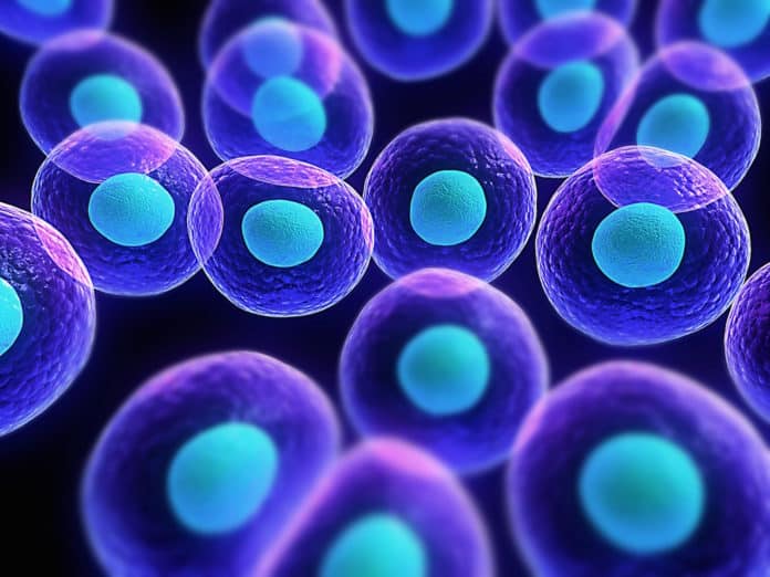
Although stem cells mimic development, a stem cell-based model of the blastocyst is lacking. The blastocyst aka the early mammalian embryo forms all embryonic as well as extra-embryonic tissues- including the placenta. Its structure consists of a spherical thin-walled layer, known as the trophectoderm, that surrounds a fluid-filled cavity sheltering the embryonic cells.
From mouse blastocysts, it is possible to derive both trophoblast and embryonic stem-cell lines, which are in vitro analogues of the trophectoderm and embryonic compartments, respectively.
Now, scientists at the Maastricht University in the Netherlands have used trophoblast and embryonic stem cells, coaxed them to cooperate trophoblast and embryonic stem cells.
“This is the first time we have created structures in the lab from stem cells which have the potential to form the whole organism – the baby, placenta and yolk sac,” Leader of the scientific team Dr. Nicolas Rivron says. For those fearing this may also result in human cloning, rest assured.
Rivron insists his research simply “helps to understand the perfect path an early embryo must take for a healthy development.” He explained that his work revealed that “it is the embryonic cells that instruct the placental cells how to organise and to implant in utero.”
“By understanding this molecular conversation, we open new perspectives to solve problems of infertility, contraception, or the adult diseases that are initiated by small flaws in the embryo,” explained Rivron. Examples of conditions that can potentially be cured by the new findings include diabetes and cardiovascular diseases.
First things first- all embryos, normally begin as blastocysts— an internal cavity that contains a small cluster of embryonic stem cells, and an outer layer of stem cells called trophoblasts. These inner embryonic cells form the embryo, while the trophoblasts eventually morph into the protective placenta that surrounds this embryo.
Now, coming to mammals, a blastocyst, which is a hollow sphere made out of fewer than 100 cells forms days after an egg is fertilized. Once this blastocyst is implanted in a uterus, the cells that form the sphere becomes the placenta, while the cells inside the blastocyst becomes the embryo.
The researchers from the present study have managed to successfully creat an artificial embryo that incorporates allows the cells to naturally intermingle, including the placenta cells.
When they implanted their model into mice, cells were seen to divide and fuse with the mother’s blood vessels, inciting pregnancy just like a natural embryo would. And to create this “blastoid”, they grew the inner and outer stem cells separately; and later introduced them within a mixture of molecules that encouraged communication and mangling.
For the first time, it is now possible to form early model embryos in unlimited numbers that implant in utero. Prof. dr. Niels Geijsen, principal investigator at the Hubrecht Institute: ‘We now have a new way to study the earliest stages of embryonic development, and to explore the influence of environmental factors on development and disease.’
Prof. dr. Clemens van Blitterswijk, department chair at the MERLN Institute of Maastricht University: ‘This research opens the path to a new biomedical discipline. We can create large numbers of model embryos and build up new knowledge by systematically testing new medical techniques and potential medicines. It also dramatically reduces the need for animal experimentation’.

