“Butterfly Eye” Camera to Aid Navigation and Identification of Cancer Cells
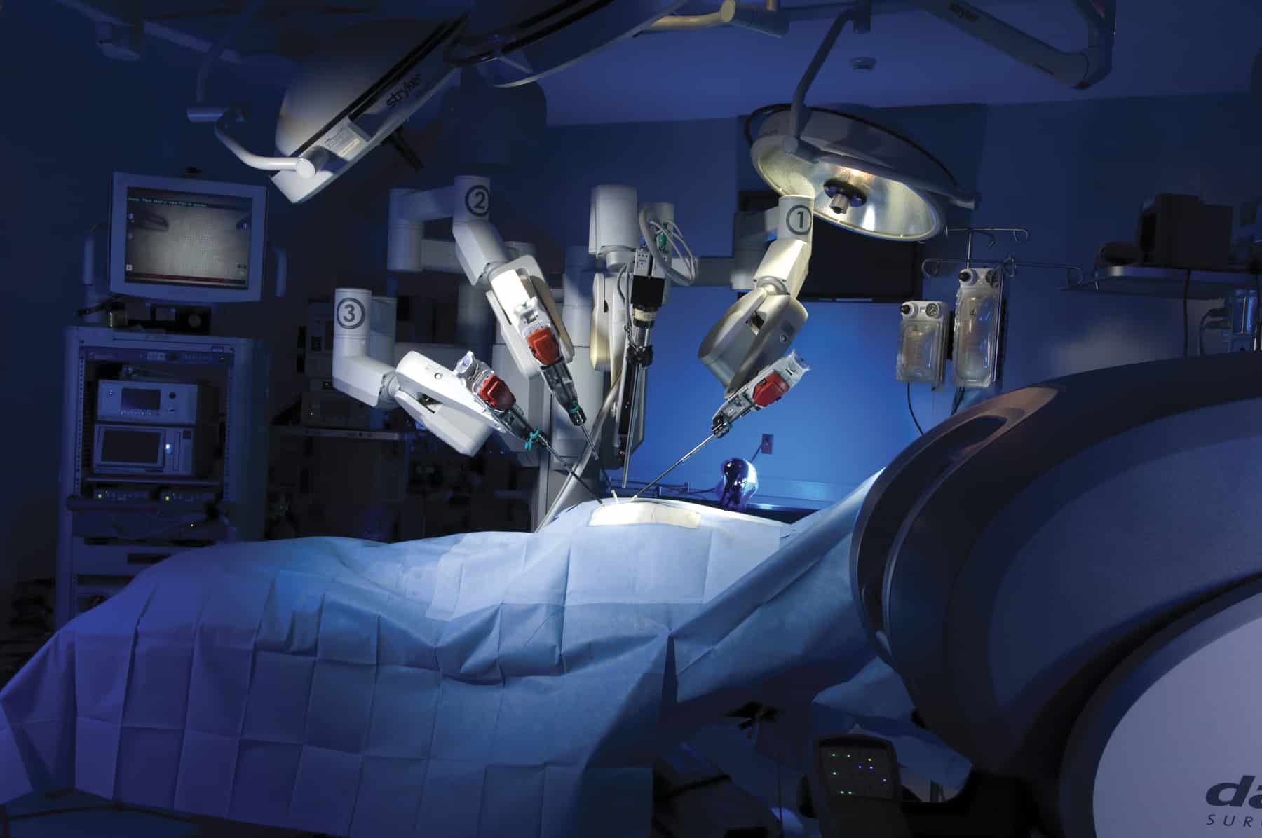
Image-guided surgery can enhance cancer treatment by decreasing, and ideally eliminating, positive tumor margins and iatrogenic damage to healthy tissue. Now, researchers at the University of Illinois at Urbana-Champaign and Washington University in St. Louis have developed a camera, inspired by the intricate visual system of a butterfly, that provides surgeons with both a traditional color image as well as a near-infrared image that makes fluorescently labeled cancerous cells visible even under bright surgical lighting.
The new camera is designed to help surgeons remove all the cancerous cells without damaging healthy tissue, making it less likely that the cancer will spread and reducing the need for multiple surgeries.
“Instead of putting together commercially available optics and sensors to build a camera for image-guided surgery, we looked to nature’s visual systems for inspiration,” said research team leader Viktor Gruev from the University of Illinois at Urbana-Champaign. “The morpho butterfly, whose eyes contain nanostructures that sense multispectral information, can acquire both near-infrared and color information simultaneously.”
“During surgery, it is imperative that all the cancerous tissue is removed, and we’ve created an imaging platform that could help surgeons do this in any hospital around the world because it is small, compact and inexpensive,” said Gruev. “Although we’ve addressed the instrumentation side, fluorescent markers targeted for cancer and approved for use in people are needed for our technology to find widespread application. Several of these are in clinical trials now, so we should see progress in this area soon.”

Many surgeons rely on sight and touch to find cancerous tissue during surgery, Gruev said. Large hospitals or cancer treatment centers may also use experimental near-infrared fluorescent agents that bind to tumors so that the surgeons can see them on specialized displays.
However, these machines are costly, making them difficult for smaller hospitals to procure; very large, making them difficult to fit into an operating suite and integrate smoothly into surgery; and require the lights to be dimmed so that the instruments can pick up the weak fluorescent signal, making it difficult for the surgeons to see.
Missael Garcia, a post-doctoral researcher at the University of Illinois at Urbana-Champaign and lead author of the paper, said they realized these problems could be mitigated by using nanostructures that resemble those of the morpho butterfly.

“Their compound eyes contain photoreceptors located next to each other such that each photoreceptor senses different wavelengths of light in a way that is intrinsically coregistered,” Garcia said in the statement.
Besides eliminating the size and light limitations of current instruments, Gruev estimates that, once mass produced, the camera and goggles will be available for $200, compared to $20,000 for existing options.
Gruev further notes that in addition to being more sensitive and accurate, it is also much smaller and lower-cost than currently available instruments that are FDA-approved.
Prototypes have been used successfully for surgery on mice, and to remove breast cancer in humans without taking extra tissue. Gruev also demonstrated his invention’s versatility by making a dye used to identify lymph nodes for biopsies to glow in the same infrared frequencies. The technique not only enabled the surgeons to find the lymph nodes more quickly but in two patients, it located nodes the surgeons would otherwise have missed.
“We showed that under bright surgical lights, our instrument was 1000 times more sensitive to fluorescence than the imagers currently approved for infrared image-guided surgery,” said Gruev. “Because the bioinspired imager can reveal fluorescence that is deep in the tissue, it sped up the process of lymph node identification and helped surgeons find lymph nodes that couldn’t be seen by eyesight alone.”
According to the researchers, the bioinspired imager would be useful for removing various types of cancers, including melanomas, prostate cancer and head and neck cancers. Because of its small size it could also be integrated into an endoscope to look for cancer during a colonoscopy, for example.