


OPTIclear: A Stunning 3D Tech that Allows for High-Resolution, Imaging
The brain is arguably the most complex organ in the human body. For centuries, efforts have been made to try and understand the structural and functional connections within the brain.
At the macroscopic level, diffusion tensor imaging and functional magnetic resonance imaging are beginning to unravel some of the complex connections between different anatomical regions. In order to better understand the workings of the organ, scientists traditionally cut into the brain and make several thin slices, later tracing the cut nerve fibres over many sections.
However, this approach is difficult and labour-intensive as the neuronal circuitries span across great distances in three dimensions (3D) and are tightly entangled microscopically. Also, due to the spatial resolution of these imaging modalities, they lack the ability to reveal details at the microcircuit or cellular level.
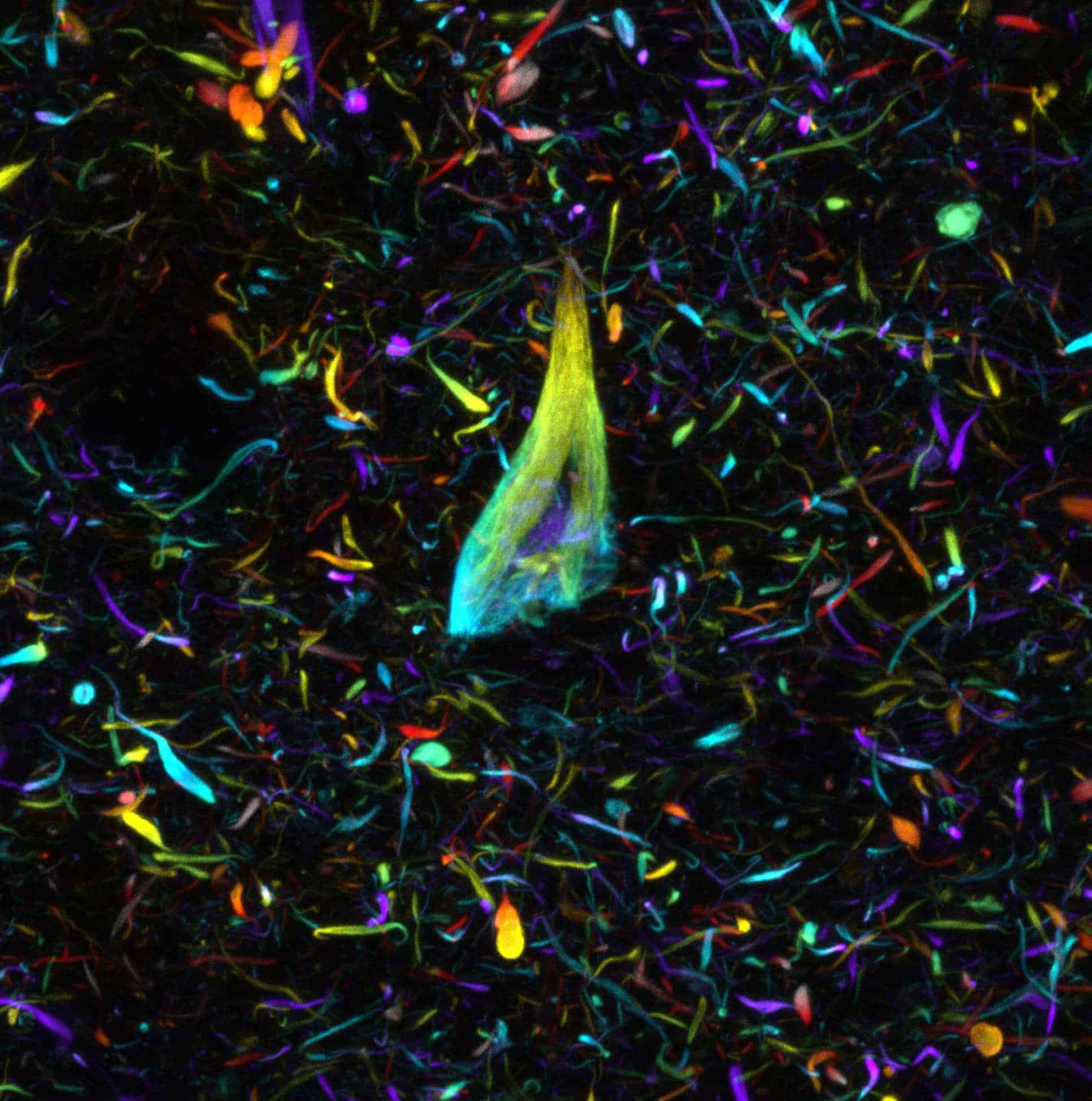
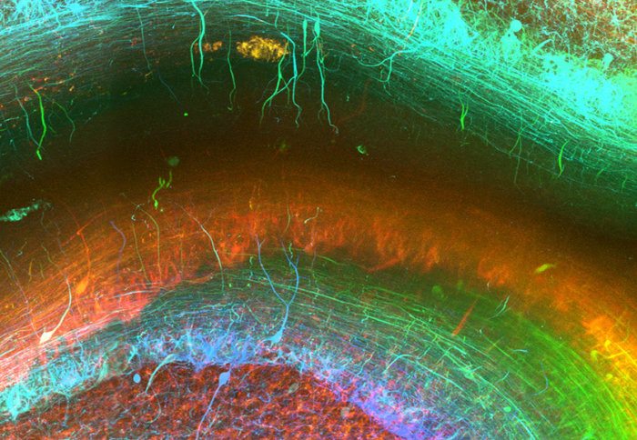
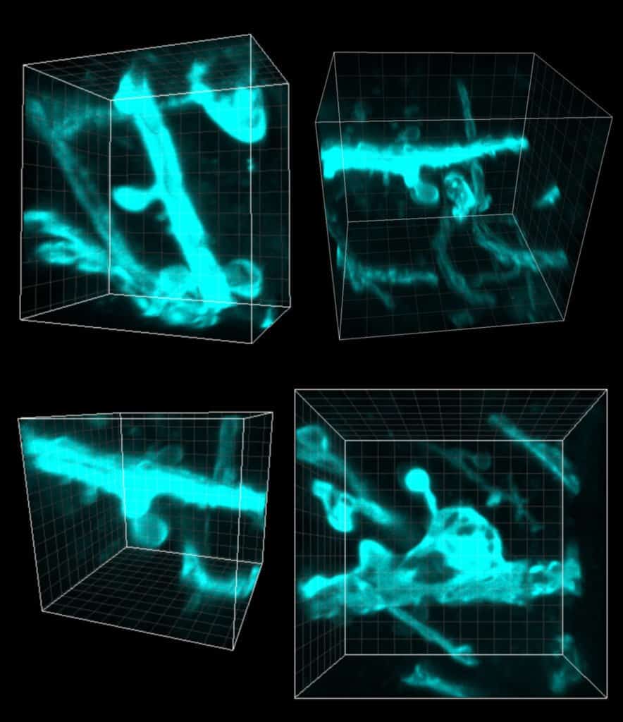
Now, developed through an international collaboration between scientists at Imperial College London and The University of Hong Kong, is a next-gen technique that enables researchers to generate 3D images of fresh and archived brain tissue samples, resulting in stunning images of the human brain at the microscopic level.
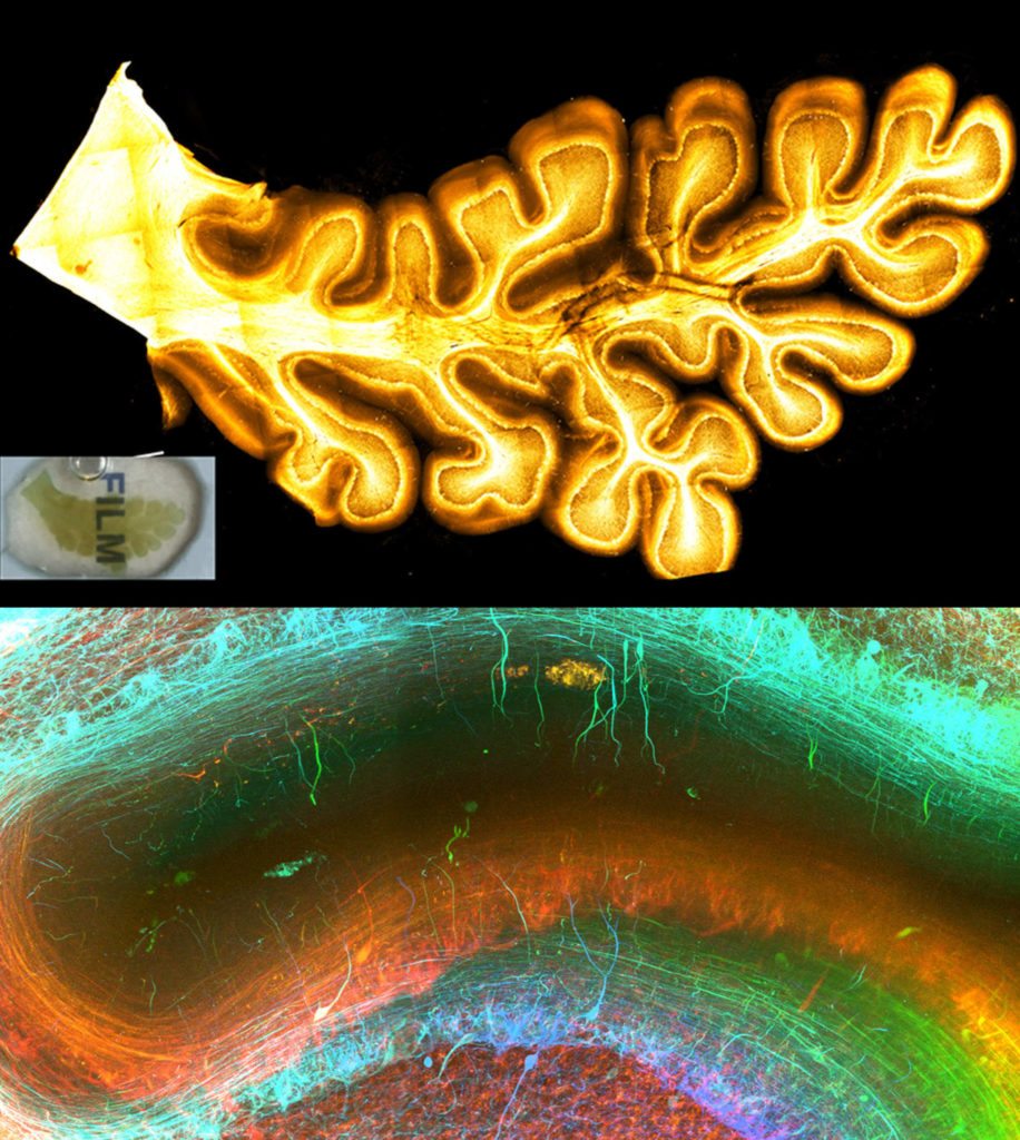
OPTIClear selectively adjusts the optical properties of tissue without damaging or changing their structural components. Combined with fluorescent staining and other tissue processing methods, the team created a simple yet versatile tool for the study of microscopic structures in the human brain.
The team performed 3D morphological analysis on human brainstem dopaminergic neurons in the millimetre scale and imaged more than 3,000 large neurons in the human basal forebrain in merely five days, a normally extremely laborious task that takes at least three weeks. These neurons have been implicated in neurological and psychiatric diseases such as dementia and depression. The researchers noted that OPTIClear can also be applied in both archived (>30 years) and clinical specimens.
“We hope that a better understanding of the connections and circuitries of the brain will help uncover the pathologies that underlie the common degenerative diseases of the brain, such as Alzheimer’s and Parkinson’s disease,” commented Prof Wutian Wu of the School of Biomedical Sciences, Li Ka Shing Faculty of Medicine, HKU, co-supervisor of the study. Lead researcher Lai Hei-Ming added: “In principle, this method is also applicable to other human organs and clinical specimens. We hope that this technique can also be used in studying other diseases, and eventually help us to unravel the mysteries of the human body.”
“We hope that a better understanding of the connections and circuitries of the brain will help uncover the pathologies that underlie the common degenerative diseases of the brain, such as Alzheimer’s and Parkinson’s disease,” commented Prof Wutian Wu from Hong Kong University.
The research team behind OPTIclear report that the method is applicable to other human organs, where it could aid in the study of other diseases at new levels of detail and “help unravel the mysteries of the human body.”


