


")

AI-Aided Microscope Boosts Malaria Diagnosis- Latest News
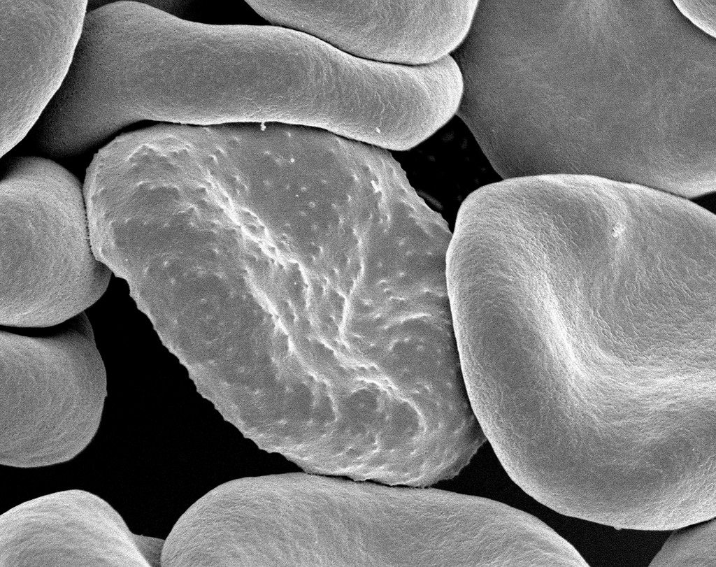
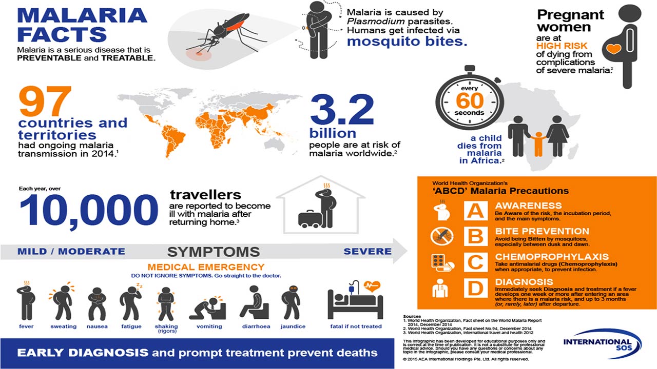
Half of the world’s population are at risk for contracting malaria, with an estimated 212 million cases in 2015. A majority of the 429,000 deaths from malaria in 2015, mostly of young children, are attributable to P. falciparum. Four other Plasmodium species—P. vivax, P. ovale, P. malarie, and, rarely, P. knowlesi—also infect humans.
Automated detection of malaria in field-prepared blood films is a challenging computer vision task with potential benefit for millions of people.
Microscopy continues to be regarded as a standard for malaria diagnosis and quantitation, in part because it can be used to detect other infectious diseases, has low incremental cost, is widely available, can measure parasite density, and can identify malaria species. Microscopy can detect low density infections if enough blood is scanned, but this is time-consuming, difficult, and tedious due to the low density and small size of parasites as well as the abundance of similar non-parasite objects.
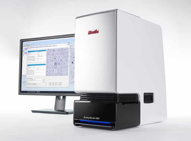
Now, Chinese manufacturer Motic, has developed an AI powered microscope that has the capability to automatically and accurately quantify malaria parasites in a blood sample.
A major difficulty with using microscopy in drug efficacy monitoring is the shortage of trained experts in regions where malaria is endemic. Therefore, the development of a computer vision system to aid in malaria diagnosis and quantitation is an appealing research goal, both because of the difficulty of the task and the high potential benefit.
Premiered at the International Conference on Computer Vision in October, the AI equipped microscope can quantify malaria parasites on par with experts, surpassing the capabilities necessary to be certified by the World Health Organization for Competency 1 microscope.
“Our goal in integrating Global Good’s advanced software into Motic’s high-quality, affordable digital slide scanner is to simplify and standardize malaria detection,” said Richard Yeung, Vice President of Motic China.
EasyScan GO works through a combination of digital slide scanning and a diagnostic software module based on machine learning and neural networking. The machine was trained by feeding it thousands of blood smear slides annotated by experts. These images were processed through a machine learning algorithm, followed by a field tests which was published and premiered at the Conference on Computer Vision.
“Malaria is one of the hardest diseases to identify on a microscope slide,” said David Bell, Director of Global Health Technologies supporting Global Good. “By putting machine learning-enabled microscopes in the hands of laboratory technicians, we can overcome two major barriers to combating the mutating parasite—improving diagnosis in case management and standardizing detection across geographies and time.”
