Alternate Neural Network That Allows For “Breathing” After Spine Injury
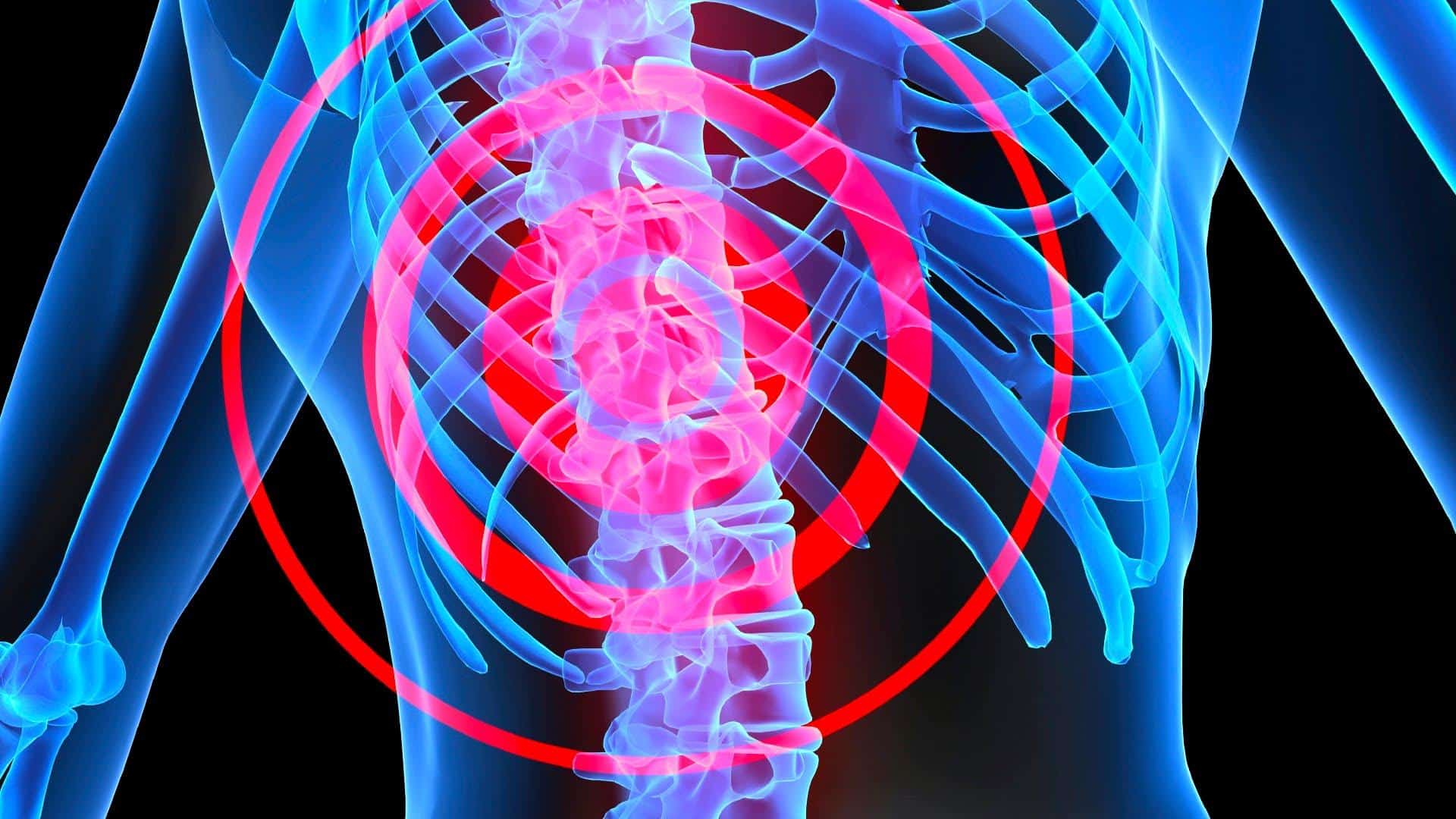
A traumatic spinal cord injury may happen because of a sudden blow or cut to the spine. A spinal cord injury often causes permanent loss of strength, sensation, and function below the site of the injury. One of the most severe consequences of spinal cord injury in the neck is losing the ability to control the diaphragm and breathe on one’s own.
Now, neuroscientists at the Case Western Reserve University School of Medicine suggest that a neural network they discovered may help restore diaphragm function after a spinal cord injury, by allowing it to contract without input from the brain.
“Respiratory signals originate in the brain and are relayed to motor neurons in the spinal cord, which then allows the diaphragm to contract. These signals are cut off after cervical spinal cord injury,” says Jared Cregg, Neurosciences graduate student at Case Western Reserve University School of Medicine in Cleveland, one of the authors of the study.
Even though this is an exciting find, scientists hope that their findings ultimately help free patients from ventilators. The pioneering work, in Cell Reports, suggests the brain may not be needed for respiration if a nerve pathway in the spine can be awakened.
The scientists restored 80 to 100 percent of breathing ability—”a remarkable return of function,” in the words of neuroscientist Jerry Silver, Ph.D., who for 30 years has worked to bring back function to the nearly 1.2 million people with spinal cord injury.
“We found a way to make the diaphragm work again in mice and hope the same approach could be applied to humans,” Jared Cregg. “We discovered a new circuit in the spinal cord that nobody knew of before. We describe it functionally based on the types of neurons in the network. We induced cervical level injury in rats and mice. We induced paralysis on the left side and found that correlated to the diaphragm function on the left side. We applied drugs that block inhibitions which kind of release the brakes so the diaphragm could start moving again.”
The researchers used a drug and a light therapy known as optogenetics to dial up this spinal system.
It appeared to control the body’s main muscle of respiration – the diaphragm, a dome-shaped sheet of muscle that sits underneath the lungs, separating the chest from the abdomen. The live adult rats that they studied had severed spinal cords, meaning the brain could not be the source of the diaphragm movement or breathing that the researchers saw after they administered the therapy.
 “We have discovered a way that may allow animals to breathe in a model of quadriplegia without the need for a respirator,” Cregg said. The new study showed quadriplegic rodents—those who underwent complete cervical spinal cord injury—had no spontaneous electrical impulses below the injury site. But by treating the rodents with pharmacologic agents, the researchers were able to induce “bursts” of electricity and how they originated in the spinal cord—not the brain. The researchers then used a laboratory technique called optogenetics to harness the impulses and induce electrical signals in the diaphragm. Optogenetics uses light to flip switches inside neurons, allowing researchers to turn on and off specific portions of the nervous system.
In neonatal mouse experiments, C1 spinal cord injuries eliminated brain-derived respiration. But, the researchers discovered electrical signals could still be transmitted from the spinal cord to the diaphragm in these mice. Even with severe spinal cord injury, the mice could maintain intermittent electrical bursts in their diaphragms consistent with breathing patterns. The findings show that the diaphragm can operate via nerve circuitry entirely separate from the brain.
They believe the treatment works by stopping other nerve signals that would normally silence the spinal system that they found. Dr. Silver said: “This is a primitive response that has been kept in the spinal cord for emergencies, like gasping and screaming in response to danger.”
Said Cregg, “Our results unexpectedly showed that diaphragm motor neurons can be controlled by two independent networks—the classical breathing network in the brain, and a spinal cord network we identify for the first time. Importantly, while previous studies hypothesized that these were parts of the same network, we show that they act completely independently.”
The newly discovered network could help spinal cord injury patients bypass missing brain signals and restore motor function below injury sites. This could include diaphragm function, to ultimately reduce their reliance on ventilators. Although the researchers say the movements they saw resembled breathing, it’s not clear yet if it would be enough to sustain life. They plan more animal studies to check.
“Our technology is still far out in terms of developing a corollary approach in humans, as our experiments indicating that we can control diaphragm motor output after cervical spinal cord injury was performed in rodent models. The technology will still need to undergo a lot of future development before it could ever be implemented as an approach to solving the human condition,” Cregg cautioned.
Said the authors, “Those attempting to enhance regeneration of [nerve cells] and restoration of a ‘simple’ motor behavior may need to consider the dynamic interplay between intact networks and networks that undergo dramatic reorganization caudal to a spinal lesion.”