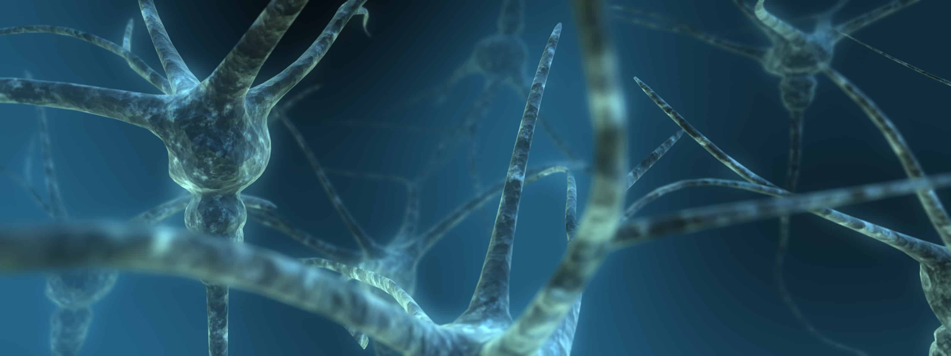
The spine is the point of connection between the brain, sensory neurons (which transmit sensory information such as temperature, pain, and touch), and motor neurons (which control the muscles). Therefore, traumatic injuries to the spine are highly complicated and can tear both motor and sensory roots — when they do, the brain loses control of the connected neurons.
Until now, surgeons used a simple method of implanting new motor roots where they were torn, prompting them to reconnect. However, this kind of repair for sensory roots has been more problematic — that is, until this recently developed procedure, in which the original sensory nerve cells are cut and removed from the root and the remaining root itself is placed directly into the spinal cord in a deeper structure.
“Doctors previously considered this type of spinal cord injury impossible to repair,” says Nicholas James, a researcher at King’s College London. “These torn root injuries can cause serious disability and excruciating pain.”
Now, scientists from the King’s College London in the UK have pinned down how one of the most cutting edge techniques works, and in particular how the body can repair itself with a little prompting from surgeons.
The team focussed on reconnecting sensory neurons to the spinal cord after traumatic injuries, looking at how the repair happens on a cellular level, and the way in which small neural offshoots grow to reconnect broken circuits in the body. Now, by recreating the technique in rats, they have new insights into the cellular processes implicated in the technique.
This new knowledge has the potential to assist the development of novel therapies for other spinal cord injuries — even those in which the spinal cord is severed.
The paper appearing in the journal, Frontiers in Neurology, details how the investigators severed the spinal cords of rats and used the new technique to reattach the sensory roots. Between 12 to 16 weeks after surgery, the researchers tested the neurons with electricity to assess whether they formed a complete neural circuit. They also analyzed the rats’ neural tissue microscopically. The electrical tests were positive, revealing a complete neural circuit and an integrated root.
The microscopic analysis of the area showed that small neural offshoots sprouted from the dendrites at the end of neurons inside the dorsal horn and spread into the implanted sensory root, resulting in a complete, functional neural circuit.
“The strategy of encouraging new growth from spinal neurons could potentially be of use in other injuries of the nervous system,” says Thomas Carlstedt, part of the team at King’s College London.
All said, although Carlstedt’s idea is exciting, what is to be noted here is the harmony between research and our body’s system- working together to repair the injuries.