


")
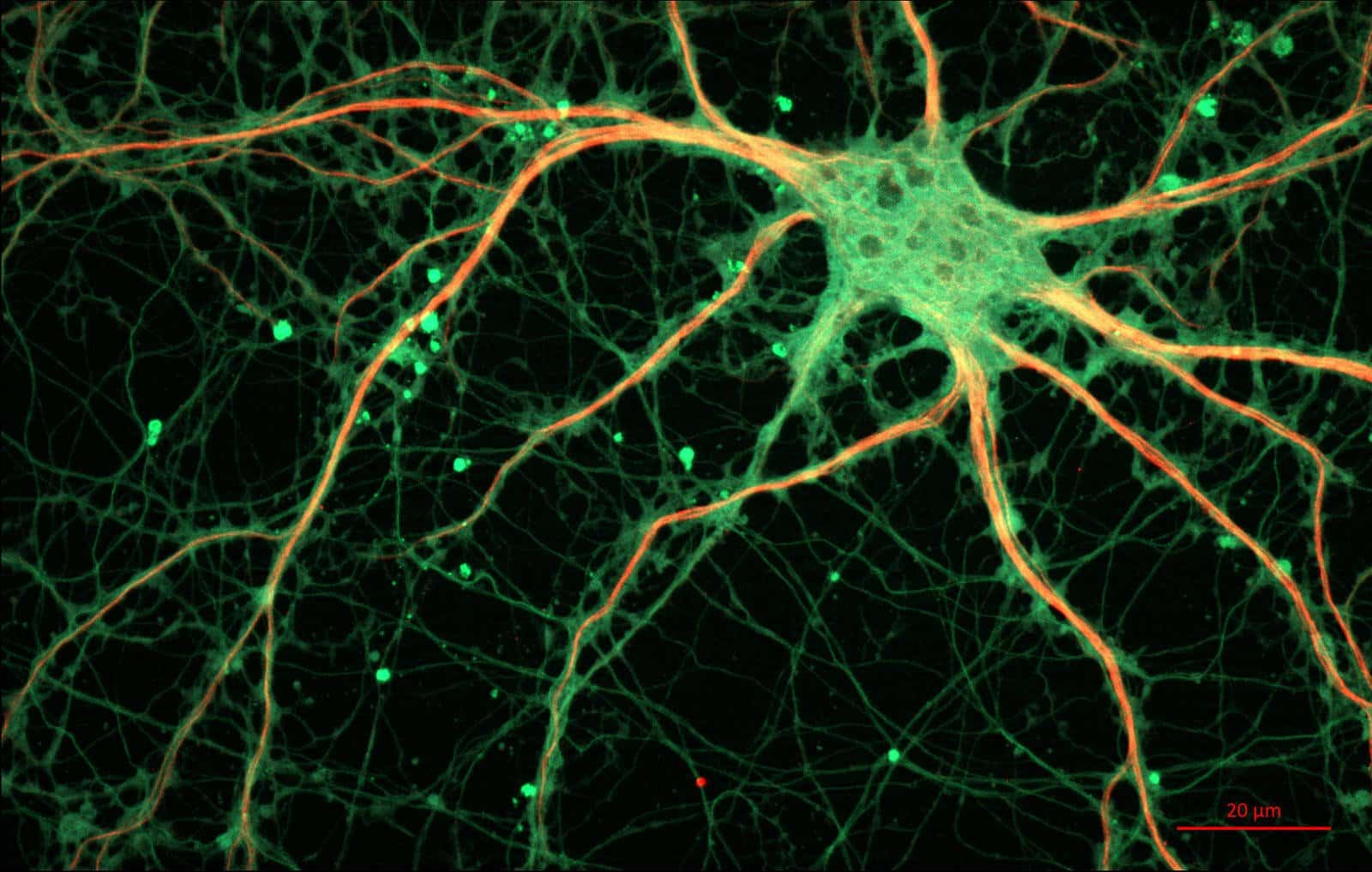
Neurons are the brain cells that store and process information. There are about 100 billion neurons in an adult human brain and we have for long known they don’t all look the same. Additionally, they are extremely difficult to differentiate even with the help of a microscope.
Therefore, scientists have turned to molecular methods in order to try and identify groups of neurons with different functions.
Now, a team from the Salk Institute for Biological Studies and the University of California San Diego has, for the first time, profiled chemical modifications in the DNA of individual neurons, giving the most detailed information yet on what makes one brain cell different from its neighbor.
DNA methylation is chemical addition of methyl groups to the bases in a DNA molecule, which alters how genes are expressed without changing their sequence. And this analysis of a neuron’s methylome, or pattern of methylated DNA, or this epigenetic change, is what has enabled the scientists to sort the neurons into subtypes and create new kinds of brain maps based on a neuron’s gene expression. The study also identifies new subtypes of neurons.
“Our research shows that we can clearly define neuronal types based on their methylomes,” said Margarita Behrens, the paper’s senior co-author. “This opens up the possibility of understanding what makes two neurons – that sit in the same brain region and otherwise look similar – behave differently.”
Single-cell methylomics is a way of profiling chemical modifications of DNA molecules in individual neurons. Each cell’s methylome—the pattern of chemical markers made up of methyl groups that stud its DNA—give a distinct readout of how its genetic switches are set.
The team studied neurons in both a mouse and a human. They used this very single-cell methylome sequencing method, applying it to 3,377 neurons from the frontal cortex of a young adult mouse and 2,784 from the frontal cortex of a deceased human male aged 25. Unlike other cells in the body, neurons have two types of methylation, so the team’s approach mapped both types.
Neurons from the mouse frontal cortex, they found, clustered into 16 subtypes based on methylation patterns, while neurons from the human frontal cortex were more diverse and formed 21 subtypes. The methylation patterns of inhibitory neurons – those that provide the brain’s “stop” signals – were more alike between mice and humans than those of the excitatory (or “go”) neurons. The study defines new subtypes of human neurons.
The team’s results add to the “brain atlas in an objective, data-driven way,” as it could lead to better understanding not only of the differences between the brains of humans and other animals but also to better understanding of brain development and dysfunction.
“This study opens a new window into the incredible diversity of brain cells,” commented Eran Mukamel, Ph.D., of the UC San Diego Department of Cognitive Science, a co-senior author of the work.
“If there’s a defect in just one percent of cells, we should be able to see it with this method,” the paper’s senior co-author, Joseph Ecker said, “Until now, we would have had no chance of picking something up in that small a percentage of cells.”
Next, the researchers plan to expand their methylome study to look at more parts of the brain, and more brains.


