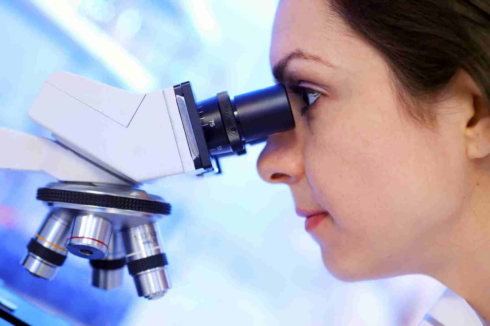
For over a century now, pathological studies have required conventional light microscopes to examine biopsy samples. However, fine-scale details of cells cannot be seen with these instruments.
Now, a team of researchers from MIT and Harvard Medical School have developed a new technique known as expansion microscopy that allows the examiner to expand a tissue sample to 100 times its original volume before imaging it which a conventional electron microscope cannot provide.
This tissue enlargement allows researchers to obtain images with a resolution of around 70 nanometers, which was previously possible only with very specialized and expensive microscopes.
In a paper published this month on the Nature Biotechnology, the study’s lead scientist, Edward Boyden and his team describe how this new technique is capable of distinguishing early-stage breast lesions with high or low risk of progressing to cancer—a task that is challenging for human observers.
“It’s a technique that could have very broad application,” says Boyden.
“Using expansion microscopy, we are able to diagnose diseases that were previously impossible to diagnose with a conventional light microscope,” says Octavian Bucur, one of the paper’s lead authors.
The team has primarily devised a novel method to fashion the samples into a state suitable for expansion. For instance, they remove the chemical stain or paraffin by exposing the tissues to a chemical solvent like xylene and then heat up the sample in another chemical called citrate. After which the tissues go through an expansion process similar to the original version of the technique, but with stronger digestion steps to compensate for the strong chemical fixation of the samples.
Using this approach, the team hopes to develop more precise diagnostics for many other diseases. To do that, scientists and doctors will need to analyze many more patient samples, allowing them to discover patterns that would be impossible to see otherwise.
Boyden and his colleagues run training courses several times a month at MIT, where visitors can come and watch expansion microscopy techniques, and they have made their protocols available on their website. They hope that many more people will begin using this approach to study a variety of diseases.
“Cancer biopsies are just the beginning,” Boyden says. “We have a new pipeline for taking clinical samples and expanding them, and we are finding that we can apply expansion to many different diseases. Expansion will enable computational pathology to take advantage of more information in a specimen than previously possible.”