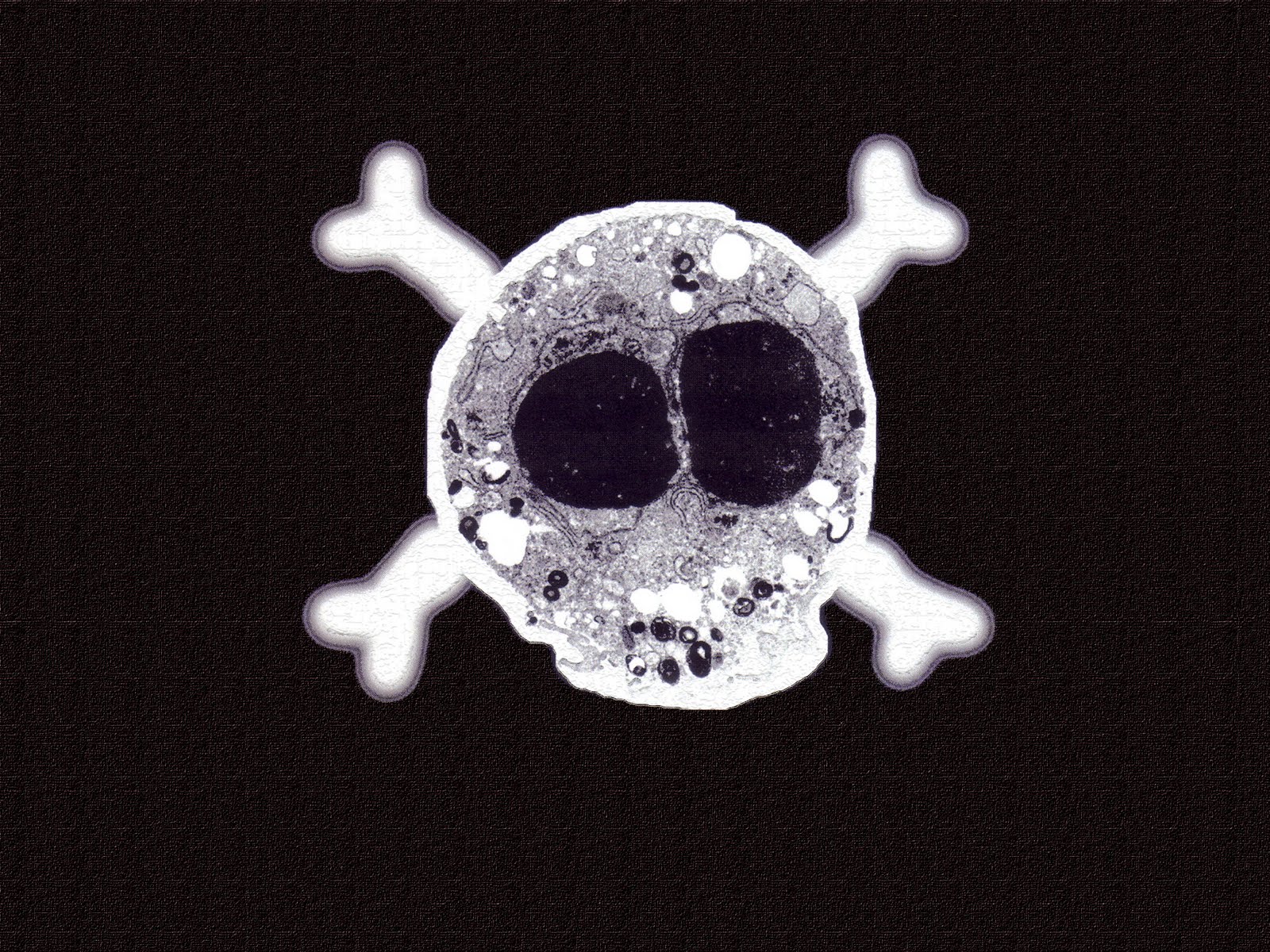
For the first time in the history of science, scientists have observed and documented the communication between dying and neighboring cells.
The cells of a multicellular organism are members of a highly organized community. The number of cells in this community is tightly regulated—not simply by controlling the rate of cell division, but also by controlling the rate of cell death.
If cells are no longer needed, they commit suicide by activating an intracellular death program, and this is called programmed cell death or more commonly, apoptosis.
Apopto
A team of scientists at the Rush University Medical Center have uncovered the system of cellular communication and its possible impact on cancer cells; they have published their findings on June 19 in an issue of The Journal Developmental Cell describing these two groundbreaking discoveries.
“I believe this discovery is going to have important ramifications for cancer biology and cancer drug development, and for the treatment of other diseases, such as diabetic foot ulcers,” said Sasha Shafikhani, PhD, associate professor in the Department of Internal Medicine at Rush Medical College, who headed the study.
Shafikhani’s team were studying pathogenic bacterium, Pseudomonas aeruginosa, to try and understand how one of the toxin it produces- ExoT, slows cell division, in addition to being capable of killing off cancer cells.
In the course of the study, particularly during a microscopic observation, they witnessed something bizarre- the dying cells released microvesicles (a type of sac) containing the protein CrkI, and these microvesicles travelled to neighboring cells and upon contact caused them to create new cells to replace the ones that are dying.
They also witnessed when they knocked out the CrkI protein during CPS, either genetically or with the ExoT toxin, they could stop cell compensatory proliferation cold. When ExoT blocked CrkI-c, the dying cells could no longer signal other cells to proliferate to replace them and this process helped Pseudomonas aeruginosa to establish their infection by ensuring damaged tissue was replaced.
Therefore, CPS was found to dog apoptosis in cancer treatment. Treated cancer cells can be induced to die, but before they do, they call on nearby cancer cells to replace them, so the cancer-fighting drug loses its effectiveness and the tumor persists.
And therapies that target both the cancer cells and nearby cells that are ready to proliferate could help stop cancer from growing and spreading.
“Knowing that it’s possible to uncouple CPS from apoptosis, we now can develop new drugs that would improve the effectiveness of treatments already in use,” Shafikhani said.


