Researchers Find New Way to Magnetise Molecules Found in the Body
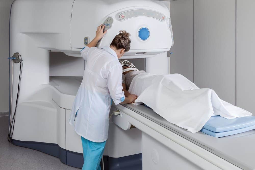
University of York researchers have now found means to shrink big, bulky hospital scanners to the size of laptop computers.
While still in the early stages, the research has made significant steps towards a new MRI method with the potential to enable doctors to personalise life-saving medical treatments and allow real-time imaging to take place in locations such as operating theatres and GP practices.
Professor Simon Duckett from the Centre for Hyperpolarisation in Magnetic Resonance at the University of York said: “What we think we have the potential to achieve with MRI what could be compared to improvements in computing power and performance over the last 40 years. While they are a vital diagnostic tool, current hospital scanners could be compared to the abacus, the recent development of more sensitive scanners takes us to Alan Turing’s computer and we are now attempting to create something scalable and low-cost that would bring us to the tablet or smartphone.”
Current MRI scanners use an extremely powerful magnet – thousands of times more powerful than a fridge magnet, for example. This changes the direction the molecules spin in the human body – polarising them to all spin in the same direction. When molecules are spinning the same way, they can be picked up by the scanner using radio waves.
The new research uses a technique that polarises glucose – and this allows much cheaper, weaker magnets to carry out the same activity. It is now theoretically possible that these magnetised, non-harmful substances could be injected into the body and visualised. Because the molecules have been hyperpolarized there would be no need to use a superconducting magnet to detect them – smaller, cheaper magnets or even just the Earth’s magnetic field would suffice.
The method, if successfully could be put into action, could enable a molecular response to be seen in real time and the low-cost, nontoxic nature of the technique would introduce the possibility of regular and repeated scans for patients. These factors would improve the ability of the medical profession to monitor and personalise treatments, possibly resulting in more successful outcomes for individuals.
“In theory, it would provide an imaging technique that could be used in an operating theatre,” added Duckett. “For example, when a surgeon extracts a brain tumour from a patient they aim to remove all the cancerous tissue while at the same time removing as little healthy tissue as possible. This technique could allow them to accurately visualise cancerous tissue at a far greater depth there and then.”
Dr Peter Rayner, Research Associate at the University of York, said: “Our method reflects one of the most significant advances in magnetic resonance in the last decade.”
Research Associate, Dr Wissam Iali added, “Given Magnetic Resonance Spectroscopy is of vital importance to the UK’s chemical and pharmaceutical industries, I see significant opportunities for them to harness our approach to improve their competitiveness.”


